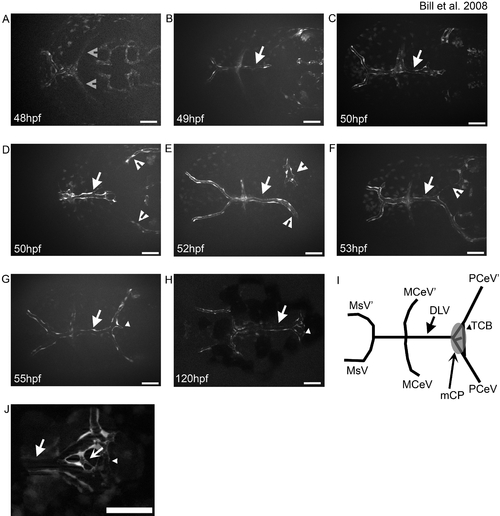
The DLV develops via angiogenic sprouting and supplies the mCP.
The development of the integral vasculature in the mCP was examined in living zebrafish larvae. The DLV (arrow in all panels) sprouts from the MCeV/MCeV′ (gray open arrowheads) via angiogenic sprouting between 40 hpf (A) and 48 hpf (B), and develops via growth cone-like filopodial extensions (C). The PCeV and PCeV′ (white open arrowheads) grow dorsomedially to meet the DLV in the roof of the fourth ventricle (D). Fusion occurs between the DLV (arrow) and PCeV or PCeV′ (E), followed by extension to the symmetric partner (PCeV or PCeV′) (F). Once connected, the DLV branches once more (G, small arrowhead) to form the trans-choroid plexus branch (TCB) (small arrowhead). By 120 hpf, the main vasculature of the mCP is in place (H). A diagrammatic representation of the final structure, with naming of the vessels and a superimposed mCP for comparison is shown (I). Small connecting vessels (concave arrow) connecting the DLV and TCB elaboration to form the mCP (J). Abbreviations: mesencephalic vein (MsV & MsV′), middle cerebral vessel (MCeV and MCeV′), dorsal longitudinal vein (DLV), posterior cerebral vein (PCeV and PCeV′) and myelencephalic choroid plexus (mCP). All images are oriented anterior to the left, and scale bars are 50 μm.
|
