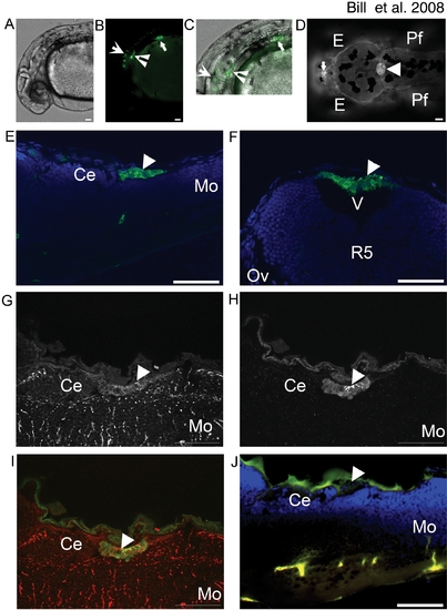
EtMn16 co-localizes with Gfap in mCP epithelia.
GFP is expressed in multiple anatomical locations in EtMn16 larvae (A–D). At 30 hpf (A–C)(lateral orientation), GFP is expressed in cells of the otic vesicle (inverted arrowhead), the pectoral fin (closed arrow), ventral hindbrain and spinal cord (open arrow) brightfield image (A), fluorescent image (B), and merged image (C). In addition to cells of the ventral hindbrain, at 4 dpf (D)(dorsal orientation), two additional structures express GFP, the diencephalic CP (dCP) (closed arrow), and the myelencephalic CP (mCP) (closed arrowhead). The mCP lies posterior to the cerebellum (Ce) [(5 dpf, 6 μm longitudinal cryosection) (E)], within the ventricle (v) dorsal to the fifth rhombomere (r5)[5 dpf, 6 μm transverse cryosection (F)]. Cells of the mCP express Gfap (G) and colocalize with GFP expressing cells (H), merged image (I) and are not seen in the negative control lacking antibodies for Gfap and GFP (J). Abbreviations: eye (E), pectoral fin (Pf), cerebellum (Ce), and medulla oblongata (Mo). All images except F are oriented anterior to the left; F is oriented dorsal to the top. Scale bar is 50 μm.
|
