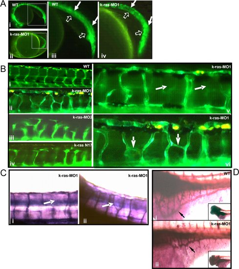
Disruption of zebrafish K-ras signaling resulted in the defective angiogenesis.
(A) Both un-injected and k-ras-MO injected fli1-GFP embryos (22 hpf) showed normal development of dorsal aorta and caudal artery (indicted by solid arrows), posterior cardinal vein and caudal vein (indicated by empty arrows). Higher magnifications of the square area in (i) and (ii) were shown in (iii) and (iv) respectively. (B) Un-injected fli1-GFP embryo at 3 dpf showed well-organized inter-segmental vessels (i), while k-ras-MO1 injected (ii, v and vi), k-ras-MO2 injected (iii) or k-rasN17 injected (iv) embryos at 3 dpf showed aberrant and irregularly organized inter-segmental vessels. (C) Alkaline phosphatase staining for k-ras-MO1 injected embryos (3 dpf) showed aberrant trunk blood vessels. (D) Alkaline phosphatase staining showed well-organized SIV (sub-intestinal vein, indicated by arrow) in wild type embryo at 3 dpf (i), while disorganized SIV (indicated by arrow) in k-ras-MO1 injected embryo (ii). Inserted figures in i and ii showed the anterior part of the embryos. All embryos shown in lateral view, with anterior to the left and dorsal to the top.
|
