Disruption of zebrafish K-ras signaling resulted in the defective hematopoiesis.
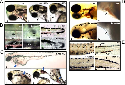
(A) k-ras-MO injected embryo showed empty heart, without or with few red blood cells inside (indicated by arrows in ii and iii), in comparison to wild type embryo, which showed plenty of red blood cells inside the heart (indicated by arrow in i). Embryos at 3 dpf (days-post fertilization). (B) Plenty of circulating red blood cells inside dorsal aorta (DA), posterior cardinal vein (PCV), inter-segmental vessels (Se), caudal artery (CA) and caudal vein (CV) in wild type embryos (i and iii), while no or less circulating red blood cells were found in k-ras-MO injected embryos inside DA, PCV, CA, CV and Se (ii and iv). Embryos at 3 dpf. (C) k-ras-MO injected embryos showed accumulated red blood cells in some sites that away from the circulation. Embryos at 3 dpf. (D) o-Dianisidine staining for wild type embryo showed hemoglobin positive cells inside branchial arches (indicated by arrow in i) and heart chambers (indicated by arrow in ii ), while k-ras-MO injected embryo showed less/negative o-Dianisidine staining for branchial arches (indicated by arrow in iii) and heart (indicated by arrow in iv). Embryos at 6 dpf. (E) o-Dianisidine staining for wild type embryo showed hemoglobin positive cells inside Se (indicated by arrows in i ), dorsal longitudinal anastomotic vessels (DLAV), CA and CV (indicated by arrows in ii), while k-ras-MO injected embryo failed to give the positive o-Dianisidine staining in these corresponding positions (indicated by arrows in iii and iv). Embryos at 6 dpf. All embryos shown are lateral view, with anterior to the left and dorsal to the top.
|
