
FIGURE
Fig. 2
- ID
- ZDB-FIG-080828-83
- Publication
- Kim et al., 2008 - Notch-regulated oligodendrocyte specification from radial glia in the spinal cord of zebrafish embryos
- Other Figures
- All Figure Page
- Back to All Figure Page
Fig. 2
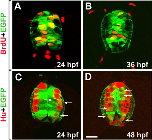
gfap+ radial glial cells divide and produce neurons. All images are transverse sections of spinal cords obtained from Tg(gfap:GFP) embryos, dorsal up. Enhanced green fluorescent protein (EGFP) fluorescence is shown as green. A,B: Bromodeoxyuridine (BrdU) labeling (red) at 24 hours postfertilization (hpf; A) and 36 hpf (B). C,D: Anti-Hu antibody labeling (red) at 24 hpf (C) and 48 hpf (D). Arrows indicate EGFP+ cells with relatively weak Hu labeling, which probably are newly born neurons. Scale bar = 20 μM. |
Expression Data
| Gene: | |
|---|---|
| Antibodies: | |
| Fish: | |
| Condition: | |
| Anatomical Terms: | |
| Stage Range: | Prim-5 to Long-pec |
Expression Detail
Antibody Labeling
Phenotype Data
Phenotype Detail
Acknowledgments
This image is the copyrighted work of the attributed author or publisher, and
ZFIN has permission only to display this image to its users.
Additional permissions should be obtained from the applicable author or publisher of the image.
Full text @ Dev. Dyn.