
Fig. 2
- ID
- ZDB-FIG-080804-22
- Publication
- Emelyanov et al., 2008 - Mifepristone-inducible LexPR system to drive and control gene expression in transgenic zebrafish
- Other Figures
- All Figure Page
- Back to All Figure Page
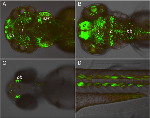
EGFP expression patterns in four independent LexPR driver-reporter lines showing different enhancer-trap events. Expression of EGFP reporter was induced in transgenic F2 embryos by adding mifepristone to the egg water at 1 μM final concentration at 24 hour post-fertilization onwards. Images were captured at 96 hour post-fertilization. No EGFP fluorescence was detected in the control populations of fish that were not treated with mifepristone (not shown). (A) Strong expression in the cell layer at the bottom of the ear (possibly sensory patches), at the olfactory bulb region (ob) and in discrete cells of midbrain (head, dorsal view). (B) Strong expression in the diencephalon (d) and cerebellum (c), lens and in discrete cells of the tectum (t) and hindbrain (hb) (head, dorsal view). (C) Expression in the olfactory bulbs (head, ventral view). (D) Specific expression in a subset of muscle cells in the somites (lateral view above yolk extension). |
Reprinted from Developmental Biology, 320(1), Emelyanov, A., and Parinov, S., Mifepristone-inducible LexPR system to drive and control gene expression in transgenic zebrafish, 113-121, Copyright (2008) with permission from Elsevier. Full text @ Dev. Biol.