
FIGURE
Fig. 3
- ID
- ZDB-FIG-080723-9
- Publication
- Shawi et al., 2008 - Identification of a BMP7 homolog in zebrafish expressed in developing organ systems
- Other Figures
- All Figure Page
- Back to All Figure Page
Fig. 3
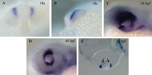
Gene expression of bmp7b in the developing eye by whole mount in situ hybridization. Dorsal (A) and lateral (B) views of an 18 somite stage embryo showing expression in the retinal neuroepithelium in the anterodorsal region of the developing optic lobe. (C) Expression remains strong in the retinal epithelium in the optic cup at 24 hpf. At 48 hpf, expression is confined in the ciliary marginal zone of the eye (arrows in E) shown as a lateral view in (D) and in a section in (E). Anterior is to the left in all panels except (A) where it is at the top. |
Expression Data
| Gene: | |
|---|---|
| Fish: | |
| Anatomical Terms: | |
| Stage Range: | 14-19 somites to Long-pec |
Expression Detail
Antibody Labeling
Phenotype Data
Phenotype Detail
Acknowledgments
This image is the copyrighted work of the attributed author or publisher, and
ZFIN has permission only to display this image to its users.
Additional permissions should be obtained from the applicable author or publisher of the image.
Reprinted from Gene expression patterns : GEP, 8(6), Shawi, M., and Serluca, F.C., Identification of a BMP7 homolog in zebrafish expressed in developing organ systems, 369-375, Copyright (2008) with permission from Elsevier. Full text @ Gene Expr. Patterns