
Fig. 5
- ID
- ZDB-FIG-080723-11
- Publication
- Shawi et al., 2008 - Identification of a BMP7 homolog in zebrafish expressed in developing organ systems
- Other Figures
- All Figure Page
- Back to All Figure Page
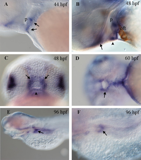
Expression in digestive system. (A) At 44 hpf, expression of bmp7b is observed in a striped pattern (arrows) in the mesoderm that is ventral to the pharynx (p). (B) This expression pattern is maintained and is more robust at 48 hpf. Embryos were co-stained with the MF20 antibody (brown) to label the myocardium as a landmark. The pharyngeal mesoderm is located immediately dorsal to the outflow tract (oft). (C) Expression is seen superficially near the developing mouth at 48 hpf. The pattern consists of three robust expression domains, one ventral (arrowhead in B and C) and symmetrical stripes on the left and right (arrows in B and C). (D) This expression domain (arrow) persists at 60 hpf although it is weaker. (E) Lateral view of a 96 hpf embryos showing expression in a restricted segment of the anterior intestinal bulb (arrow). (F) Higher magnification of embryo seen in (E). |
| Gene: | |
|---|---|
| Antibody: | |
| Fish: | |
| Anatomical Terms: | |
| Stage Range: | High-pec to Day 4 |
Reprinted from Gene expression patterns : GEP, 8(6), Shawi, M., and Serluca, F.C., Identification of a BMP7 homolog in zebrafish expressed in developing organ systems, 369-375, Copyright (2008) with permission from Elsevier. Full text @ Gene Expr. Patterns