
- Title
-
Identification of a BMP7 homolog in zebrafish expressed in developing organ systems
- Authors
- Shawi, M., and Serluca, F.C.
- Source
- Full text @ Gene Expr. Patterns
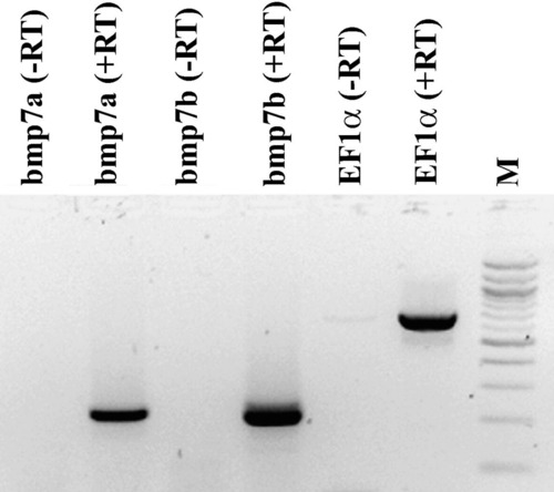
Maternal expression of zebrafish bmp7 genes. RT-PCR of elongation factor 1 alpha (Ef1α), snh/bmp7a and bmp7b from RNA extracted at stage 2–8 cells. Both bmp7 orthologs are maternally expressed. Controls omitting the reverse transcriptase step are indicated by (-RT). M: Molecular marker (100 bp ladder). |
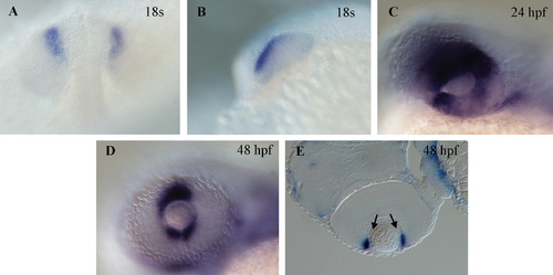
Gene expression of bmp7b in the developing eye by whole mount in situ hybridization. Dorsal (A) and lateral (B) views of an 18 somite stage embryo showing expression in the retinal neuroepithelium in the anterodorsal region of the developing optic lobe. (C) Expression remains strong in the retinal epithelium in the optic cup at 24 hpf. At 48 hpf, expression is confined in the ciliary marginal zone of the eye (arrows in E) shown as a lateral view in (D) and in a section in (E). Anterior is to the left in all panels except (A) where it is at the top. EXPRESSION / LABELING:
|
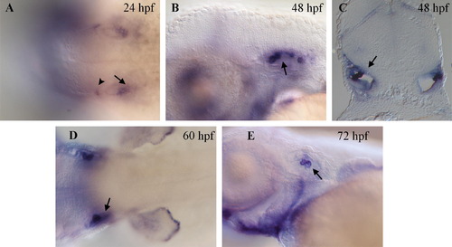
Expression of bmp7b detected by in situ hybridization in the developing ear. (A) Transcripts of bmp7b can be seen at 24 hpf in the posterior half of the otic vesicle (arrow) and at its anterior most point (arrowhead). (B) Lateral view and (C) cross sectional view of the developing ear at 48 hpf. Strong bmp7b expression can be seen in the forming semi-circular canals (arrows) and weaker expression is observed throughout the epithelium. Expression remains strong in the semi-circular canals at 60 hpf (D, arrow) and 72 hpf (E, arrow) when little or no expression remains in the epithelial component. Anterior is to the left in all panels showing whole mount embryos. EXPRESSION / LABELING:
|
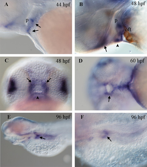
Expression in digestive system. (A) At 44 hpf, expression of bmp7b is observed in a striped pattern (arrows) in the mesoderm that is ventral to the pharynx (p). (B) This expression pattern is maintained and is more robust at 48 hpf. Embryos were co-stained with the MF20 antibody (brown) to label the myocardium as a landmark. The pharyngeal mesoderm is located immediately dorsal to the outflow tract (oft). (C) Expression is seen superficially near the developing mouth at 48 hpf. The pattern consists of three robust expression domains, one ventral (arrowhead in B and C) and symmetrical stripes on the left and right (arrows in B and C). (D) This expression domain (arrow) persists at 60 hpf although it is weaker. (E) Lateral view of a 96 hpf embryos showing expression in a restricted segment of the anterior intestinal bulb (arrow). (F) Higher magnification of embryo seen in (E). |
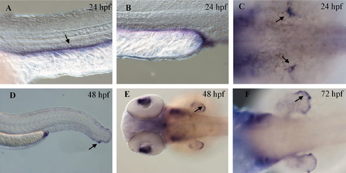
Expression in the pronephros and developing fins. (A) Bmp7b expression is seen in the pronephric duct (arrow) at 24 hpf. Strongest expression is seen at the posterior end near the cloaca (B). (C) The pectoral fin buds (arrows) also show robust expression of bmp7b at 24 hpf. (D) At 48 hpf, bmp7b is expressed at the outer edge of both ventral and dorsal fin tissue with strongest expression seen at the posterior end (arrow). Dorsal views of 48 hpf (E) and 72 hpf (F) embryos with expression of bmp7b observed at the outer ridge of the pectoral fin (arrows). EXPRESSION / LABELING:
|
Reprinted from Gene expression patterns : GEP, 8(6), Shawi, M., and Serluca, F.C., Identification of a BMP7 homolog in zebrafish expressed in developing organ systems, 369-375, Copyright (2008) with permission from Elsevier. Full text @ Gene Expr. Patterns