
Fig. S1
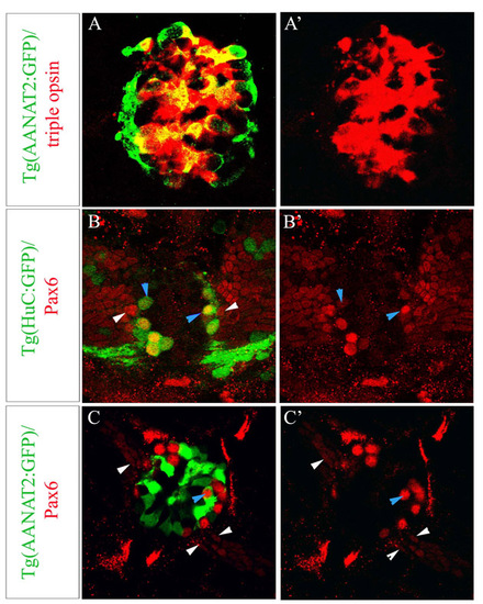
(A-A′) Optical sections of Tg(AANAT2:GFP) epiphysis at 2 days of development stained with a triple red opsin, exorhodopsin, rhodopsin probe and GFP. All opsin-positive cells are Tg(AANAT2:GFP)+; conversely, only 87.2±4.14% of Tg(AANAT2:GFP)+ cells are positive for one of the three opsins. (B-C′) Optical sections of Tg(HuC:GFP) (B,B′) and Tg(AANAT2:GFP) (C,C′) epiphyses at 48 hours labelled with a Pax6 antibody and GFP. Tg(HuC:GFP)+ cells are also positive for Pax6 (blue arrowheads). Numerous Pax6+, Tg(HuC:GFP)- neuroepithelial cells are observed in the ventrolateral part of the epiphysial area (white arrowheads). Pax6 and Tg(AANAT2:GFP) expression is mostly exclusive except for rare cells expressing both markers (blue arrowheads). |