
Fig. 3
- ID
- ZDB-FIG-080702-45
- Publication
- Hyatt et al., 1996 - Retinoic acid alters photoreceptor development in vivo
- Other Figures
- All Figure Page
- Back to All Figure Page
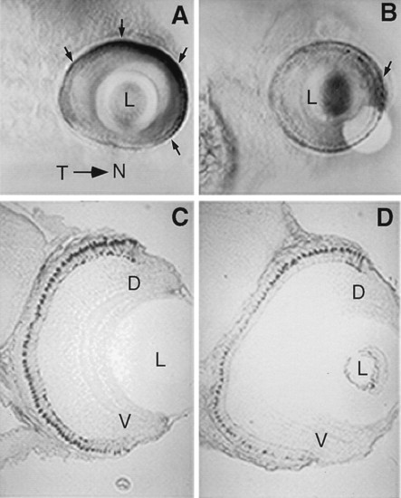
Localization of mRNA transcripts for ultraviolet opsin by whole mount in situ hybridization at day 5 pf in control embryos (A) and RA-treated embryos (B) in lateral view. (A) In controls, ultraviolet opsin expression is most robust within the nasal retina and faint expression is also observed in the temporal retina (arrows indicate regions of high expression). (B) After three days of RA treatment, ultraviolet opsin expression is significantly decreased at day 5 in RA-treated embryos throughout most of the retina. The only region of dense staining is found within the nasal retina (arrow). The localization of blue opsin transcripts by in situ hybridization at day 5 in transverse sections (5 μm) in control (C) and RA-treated (D) embryos. Blue opsin staining is generally weaker in RA-treated embryos (D). Furthermore, staining patterns of cells expressing the blue opsin appears graded in RA-treated embryos along the dorsal-ventral axis of the retina. The size of cones expressing the blue-sensitive opsin is decreased, particularly in regions ventral to the dorsal retina. D, dorsal retina; V, ventral retina; L, lens; Y, yolk. |