
Fig. 4
- ID
- ZDB-FIG-080620-53
- Publication
- Li et al., 1997 - A dominant form of inherited retinal degeneration caused by a non-photoreceptor cell-specific mutation
- Other Figures
- All Figure Page
- Back to All Figure Page
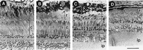
Histological sections showing the photoreceptor layer of 13-month-old wild-type and nba retinas. (A) A section from the retina of a wild-type fish. In the light adapted retina, the rods (r) sit distal to the cones (c). Processes from the pigment epithelium (PE) extend between the outer segments of the rods. (B-D) Sections from the central retina of one nba fish showing the variability of degeneration seen in various regions of the affected retina (see text for details). in, inner nuclear layer; ip, inner plexiform layer. Arrows in B indicate the large lipid droplets in PE. [Bar = ≈100 μm (A) and 80 μm (B-D).] |