
Fig. 5
- ID
- ZDB-FIG-080620-54
- Publication
- Li et al., 1997 - A dominant form of inherited retinal degeneration caused by a non-photoreceptor cell-specific mutation
- Other Figures
- All Figure Page
- Back to All Figure Page
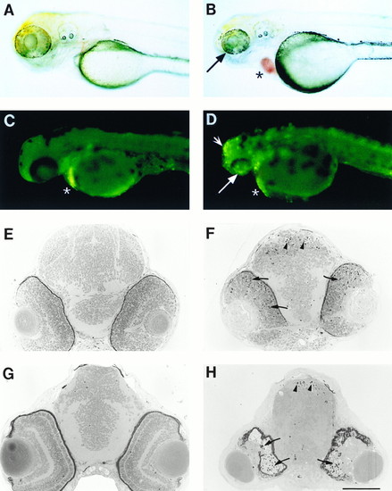
Photographs of live embryos (A-D) and histological sections of the brain and retinas (E-H) of wild-type (Left) and homozygous nba (Right) fish. (A and B) Photograph of 2.5-day-old wild-type and nba embryos. Note the smaller eye (arrow) and blood cells that have pooled in the heart (asterisk) in nba fish. (C and D) Acridine orange staining of 2.5-day-old wild-type and nba embryos. Brightly staining apoptotic cells are seen in the retina (arrow) and tectum (arrowhead) in nba fish. Asterisks indicate nonspecific staining of yolk cells. (E and F) Transverse sections through the brain and retina of 2.5-day-old wild-type and nba embryos. Note the darkly stained dying cell in both the retina (arrows) and tectum (arrowheads) in nba fish. (G and H) Transverse sections of 3.5-day-old wild-type and nba embryos. By this time most of the retinal cells have degenerated (arrows), and the tectum continuous to show evidence of cell death (arrowheads) in nba fish. [Bar = ≈250 μm (A-D) and 100 μm (E-H)]. |