FIGURE

Fig. S1

ID
ZDB-FIG-080604-76
Publication
Peri et al., 2008 - Live Imaging of Neuronal Degradation by Microglia Reveals a Role for v0-ATPase a1 in Phagosomal Fusion In Vivo
Other Figures
All Figure Page
Back to All Figure Page
Fig. S1

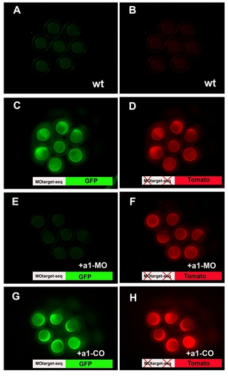
The a1MO binds to its target sequence
A-H Pools of 6-hours old zebrafish embryos. A, C, E, G Green channel. B, D, F, H Red channel. A and B uninjected embryos. C and D Embryos injected with a1- Mo target-sequence-GFP-RNA (C, green) and 5-bases-mismatck-sequence- Tomato (D, red). E-F Embryos injected with a1-Mo target-sequence-GFP-RNA (E, green) and 5-bases-mismatck-sequence-Tomato (F, red). Injection of both RNAs was followed by the injection of the a1-MO. G-H Embryos injected with a1- Mo target-sequence-GFP-RNA (G, green) and 5-bases-mismatck-sequence- Tomato (H, red). Injection of both RNAs was followed by the injection of the COa1- MO.

Expression Data

Expression Detail
Antibody Labeling
Phenotype Data

Phenotype Detail
Acknowledgments
This image is the copyrighted work of the attributed author or publisher, and ZFIN has permission only to display this image to its users. Additional permissions should be obtained from the applicable author or publisher of the image.

Reprinted from Cell, 133(5), Peri, F., and Nüsslein-Volhard, C., Live Imaging of Neuronal Degradation by Microglia Reveals a Role for v0-ATPase a1 in Phagosomal Fusion In Vivo, 916-927, Copyright (2008) with permission from Elsevier. Full text @ Cell