|
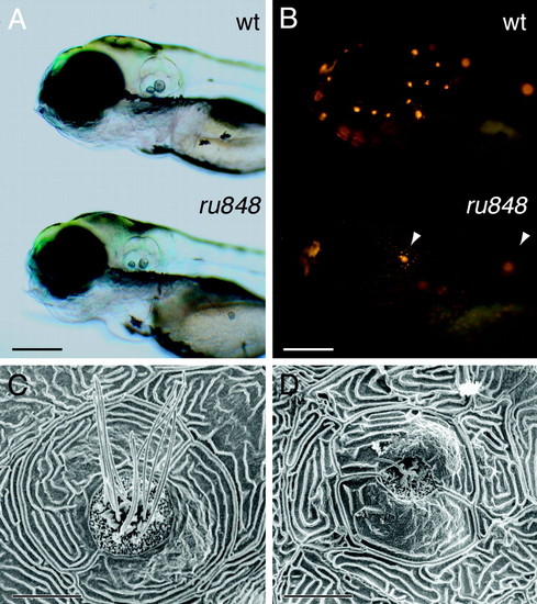
Disruption of lateral-line hair cells by the ru848 mutation. (A) Transmitted-light illumination shows the grossly normal structure of the otocysts in WT (Upper) and mutant (Lower) larvae at 5 dpf. (B) Under fluorescent illumination, the neuromasts form a characteristic pattern on the head of a WT larva (Upper) labeled with 4-Di-2-ASP. The mutant larva (Lower) bears only two brightly labeled neuromasts (arrowheads). (C) In a scanning electron micrograph, the center of a WT neuromast at 6 dpf displays hair bundles with short stereocilia and long kinocilia. The neuromast is enclosed by two periderm cells. (D) In a mutant, the periderm cells appear relatively normal, but in the center of the neuromast stand only two kinocilia that are much shorter than those seen in the WT. (Scale bars: 200 μm, A and B; 10 μm, C and D.)
|