|
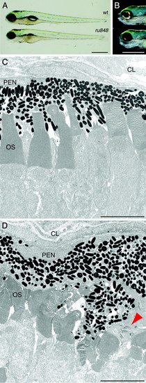
Morphology of ru848 mutant and WT larvae at 5 dpf. (A) A transilluminated mutant larva (Lower) lacks an inflated swim bladder and has a slightly smaller eye than the WT (Upper). (B) Incident illumination shows reflective iridiphores that cover almost the entire eye in the WT larva (Upper) but are sparse in the mutant (Lower). (C) In a transmission electron micrograph, the photoreceptor outer segments (OS) in the WT retina are neatly arrayed and interdigitated with processes of the pigment-epithelial cells. (D) In the mutant retina, the outer segments are disheveled and the pigment-epithelial cells are hypertrophic and disorganized. A cluster of cytoplasmic inclusions in a mutant pigment epithelial cell is marked by a red arrowhead. CL, choroid layer; PEN, pigment epithelial nucleus. (Scale bars: 500 μm, A and B;5 μm, C and D.)
|