
- Title
-
Mutation of the zebrafish choroideremia gene encoding Rab escort protein 1 devastates hair cells
- Authors
- Starr, C.J., Kappler, J.A., Chan, D.K., Kollmar, R., and Hudspeth, A.J.
- Source
- Full text @ Proc. Natl. Acad. Sci. USA
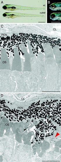
Morphology of ru848 mutant and WT larvae at 5 dpf. (A) A transilluminated mutant larva (Lower) lacks an inflated swim bladder and has a slightly smaller eye than the WT (Upper). (B) Incident illumination shows reflective iridiphores that cover almost the entire eye in the WT larva (Upper) but are sparse in the mutant (Lower). (C) In a transmission electron micrograph, the photoreceptor outer segments (OS) in the WT retina are neatly arrayed and interdigitated with processes of the pigment-epithelial cells. (D) In the mutant retina, the outer segments are disheveled and the pigment-epithelial cells are hypertrophic and disorganized. A cluster of cytoplasmic inclusions in a mutant pigment epithelial cell is marked by a red arrowhead. CL, choroid layer; PEN, pigment epithelial nucleus. (Scale bars: 500 μm, A and B;5 μm, C and D.) PHENOTYPE:
|
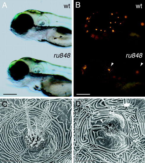
Disruption of lateral-line hair cells by the ru848 mutation. (A) Transmitted-light illumination shows the grossly normal structure of the otocysts in WT (Upper) and mutant (Lower) larvae at 5 dpf. (B) Under fluorescent illumination, the neuromasts form a characteristic pattern on the head of a WT larva (Upper) labeled with 4-Di-2-ASP. The mutant larva (Lower) bears only two brightly labeled neuromasts (arrowheads). (C) In a scanning electron micrograph, the center of a WT neuromast at 6 dpf displays hair bundles with short stereocilia and long kinocilia. The neuromast is enclosed by two periderm cells. (D) In a mutant, the periderm cells appear relatively normal, but in the center of the neuromast stand only two kinocilia that are much shorter than those seen in the WT. (Scale bars: 200 μm, A and B; 10 μm, C and D.) |
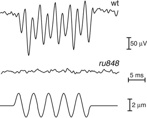
Analysis of hair-cell function in WT and mutant fish at 7 dpf. While a vibratory stimulus (200 Hz, ±2 µm; Bottom) was provided by a glass probe, extracellular microphonic potentials were recorded and averaged over 50-100 stimulus epochs. The WT larvae displayed a robust electrical response at twice the stimulus frequency (Top), reflecting the activation of two populations of hair cells with opposing orientations. No response to the vibratory stimulus was seen in the 10 mutant larvae examined (Middle). PHENOTYPE:
|
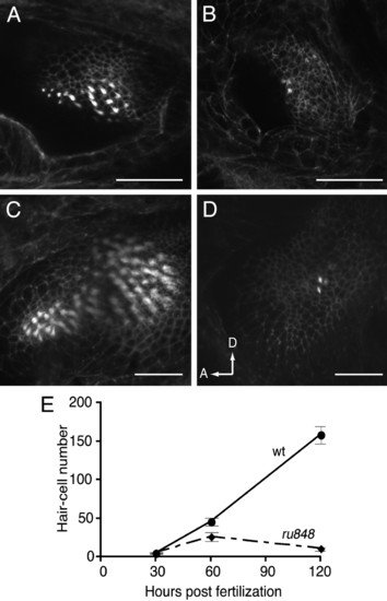
Reduction in the number of inner-ear hair cells in mutant larvae. The total number of hair cells was assessed at three developmental times by counting the number of Alexa 488-phalloidin-labeled hair bundles in all sensory epithelia. A reduction in number became noticeable from 60 hpf, as shown here in single confocal sections through WT (A) and mutant (B) posterior maculae. By 120 hpf, many more hair cells had been added to the posterior macula in the WT (C), but little change was observed in the mutant (D), as seen in projections of Z-series through the posterior maculae. In D, arrows show anterior (A) and dorsal (D) directions. (Scale bars: 50 μm.) (E) Similar results were observed when hair cells from all five sensory epithelia in the inner ear were counted at three developmental stages. At least eight larvae were examined for each data point. |
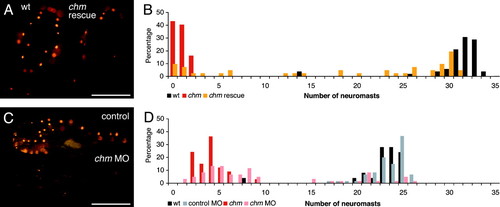
Confirmation of the mutation in chm as responsible for the ru848 phenotype. (A) REP1 mRNA transcribed in vitro rescues the mutant phenotype. Dorsal views of WT (Left) and mutant (Right) larvae at 5 dpf after injection with in vitro-transcribed REP1 mRNA. The mutant larva has been partially rescued by the exogenous transcript. On the left side of the mutant fish, an almost normal number of neuromasts can be observed, but on the right side, the only labeled neuromast is that typically seen above the ear in uninjected mutant larvae. The WT larva was also injected with the exogenous transcript but showed no sign of impairment caused by overexpression of this gene. (B) A histogram of the number of labeled neuromasts at 5 dpf in mutant larvae after injection with chm mRNA (orange) documents a distribution distinct from those for uninjected WT (black) and mutant (red) controls. The injected mutant larvae display a broad distribution in neuromast number, with the majority of the mutants displaying at least a partially rescued phenotype. (C)Ina "knockdown" of REP1 translation, the morpholino (MO)-injected larva (Lower) has only one labeled neuromast above the ear and one in the posterior lateral line. The control-injected larva (Upper) has the normal complement of neuromasts. (D) In a histogram of neuromast numbers at 3 dpf, WT larvae injected with a control morpholino (MO) (gray) have a distribution in neuromast number similar to that seen in uninjected WT larvae (black). In contrast, the majority of WT larvae injected with the chm-targeted morpholino (pink) display a strikingly reduced number of labeled neuromasts, similar to that in uninjected mutant larvae (red). (Scale bars: 500μm.) PHENOTYPE:
|

Unillustrated author statements PHENOTYPE:
|