
FIGURE
Fig. 7
Fig. 7
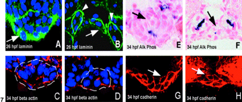
Tissue morphogenesis and cell polarization are coincident within the developing gut tube. (A, B) 26 hpf. Laminin surrounds the newly formed foregut (A, arrow) and hindgut (B, arrow) Arrowheads, pronephric ducts. (C–H) 34 hpf. Apical β-actin (C, D) and alkaline phosphatase activity (E, F) are seen in the gut for the first time at this stage (foregut: white dashed line in C, arrow in E; hindgut: white dashed line in D, arrow in F). Immunoreactive cadherin within the foregut (G arrow) and hindgut (H arrow) is also evident at 34 hpf. |
Expression Data
| Antibodies: | |
|---|---|
| Fish: | |
| Anatomical Terms: | |
| Stage Range: | Prim-5 to Prim-15 |
Expression Detail
Antibody Labeling
Phenotype Data
Phenotype Detail
Acknowledgments
This image is the copyrighted work of the attributed author or publisher, and
ZFIN has permission only to display this image to its users.
Additional permissions should be obtained from the applicable author or publisher of the image.
Reprinted from Developmental Biology, 255(1), Wallace, K.N. and Pack, M., Unique and conserved aspects of gut development in zebrafish, 12-29, Copyright (2003) with permission from Elsevier. Full text @ Dev. Biol.