
Fig. 2
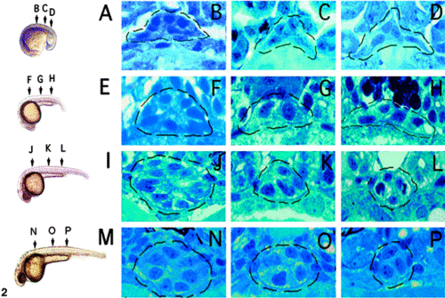
Formation of the zebrafish gut tube at mid-somite stages. (A) Lateral view of an 18-hpf embryo with corresponding histological sections (B–D). Endoderm at this stage does not appear organized. (E) Lateral view of a 21-hpf embryo with corresponding histological sections (F–H). Endoderm near the level of the finbuds (F) assumes a circular or radial organization with peripherally located nuclei. Endoderm caudal to the beginning of the yolk extension (G, H) appears unchanged from 18 hpf. (I) Lateral view of a 26-hpf embryo with corresponding histological sections (J–L). Radially organized endoderm is present rostrally from the finbuds to the yolk extension (J) and near in the common pronephric duct (L) but not within the intervening region (K). (M) Lateral view of a 34-hpf embryo. At this stage, the endoderm from the finbuds to the common pronephric duct has adopted a tube-like configuration. The gut lumen is visible at this stage (N) and also recognizable in transmission electron micrographs of 30-hpf embryos (not shown). Dashed line circles the gut endoderm in all histological sections. |
Reprinted from Developmental Biology, 255(1), Wallace, K.N. and Pack, M., Unique and conserved aspects of gut development in zebrafish, 12-29, Copyright (2003) with permission from Elsevier. Full text @ Dev. Biol.