FIGURE
Fig. 1
Fig. 1
|
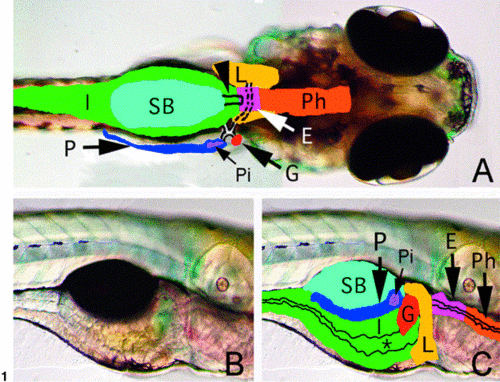
Larval zebrafish digestive system anatomy. (A) Dorsal view of a 5- dpf larva. Overlay outlines the pharynx (Ph), esophagus (E), liver (L), pancreas (P) with solitary islet (Pi), gallbladder (G), swimbladder (SB), and intestine (I). Broken and solid lines depict ducts of the liver, pancreas, and gallbladder. Swimbladder pneumatic duct is denoted by arrowhead. (B, C) Lateral view of 5-dpf larva with overlay. *, marks intestinal lumen. |
Expression Data
Expression Detail
Antibody Labeling
Phenotype Data
Phenotype Detail
Acknowledgments
This image is the copyrighted work of the attributed author or publisher, and
ZFIN has permission only to display this image to its users.
Additional permissions should be obtained from the applicable author or publisher of the image.
Reprinted from Developmental Biology, 255(1), Wallace, K.N. and Pack, M., Unique and conserved aspects of gut development in zebrafish, 12-29, Copyright (2003) with permission from Elsevier. Full text @ Dev. Biol.

