|
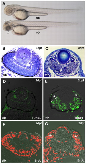
Retinal neurons undergo extensive apoptosis in the piy mutant. (A) Morphology of wild-type and piy mutant zebrafish embryos at 2 dpf. Both embryos were treated with phenyltiourea to prevent pigmentation. Extensive cell death is observed in the retina and tectum of the piy mutant (arrowheads). (B,C) Plastic sections of wild-type (B) and piy mutant (C) retinas. In the piy mutant retinas, extensive cell death occurs in the central retina, where differentiated neurons are normally located. Retinal stem cells are retained in the CMZ of the piy mutant (asterisks). (D,E) TUNEL of wild-type (D) and piy mutant (E) retinas. In the piy mutant, most cells in the central retina are TUNEL-positive (green), whereas there are only a few TUNEL-positive cells in the CMZ (asterisks). (F,G) BrdU labeling of wild-type (F) and piy mutant (G) retinas. In the piy mutant, retinal stem cells in the CMZ incorporate BrdU (asterisks).
|