
Fig. 4
- ID
- ZDB-FIG-080403-5
- Publication
- Xing et al., 2008 - Crystal Structure of a Full-Length beta-Catenin
- Other Figures
- All Figure Page
- Back to All Figure Page
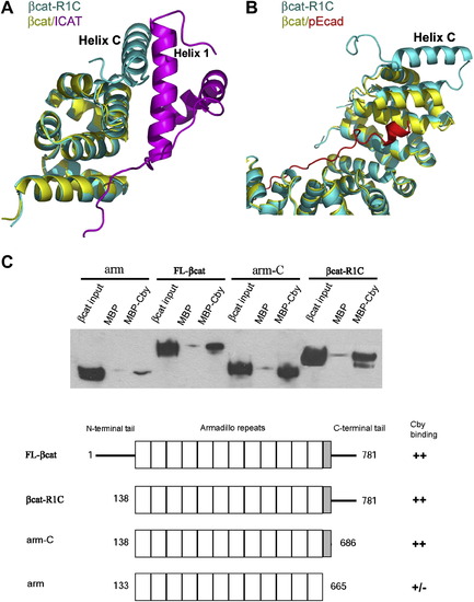
The Role of Helix C in β-Catenin Protein-Protein Interactions (A) Potential interactions between β-catenin helix C and ICAT. β-catenin-R1C (βcat-R1C, cyan) is superimposed onto β-catenin/ICAT (βcat/ICAT, yellow and magenta), based on the Cα's of β-catenin armadillo repeats 10–12 (residues 563–663). (B) Helix C is unlikely to affect the interaction between the phophorylated E-cadherin and β-catenin armadillo repeats. β-catenin-R1C (βcat-R1C, cyane) is superimposed with β-catenin/phospho-Ecadherin (βcat/pEcad, yellow and red, PDB code: 1I7W) based on all twelve armadillo repeats. (C) The helix C is required for β-catenin to interact with Chibby. Purified MBP-Chibby protein was immobilized to amylose beads; purified β-catenin and β-catenin fragments were tested for their binding to immobilized MBP-Chibby. The total input and bound β-catenin and β-catenin fragments were visualized by Western blot. |
Reprinted from Structure (London, England : 1993), 16(3), Xing, Y., Takemaru, K., Liu, J., Berndt, J.D., Zheng, J.J., Moon, R.T., and Xu, W., Crystal Structure of a Full-Length beta-Catenin, 478-487, Copyright (2008) with permission from Elsevier. Full text @ Structure