
Fig. 3
- ID
- ZDB-FIG-080403-4
- Publication
- Xing et al., 2008 - Crystal Structure of a Full-Length beta-Catenin
- Other Figures
- All Figure Page
- Back to All Figure Page
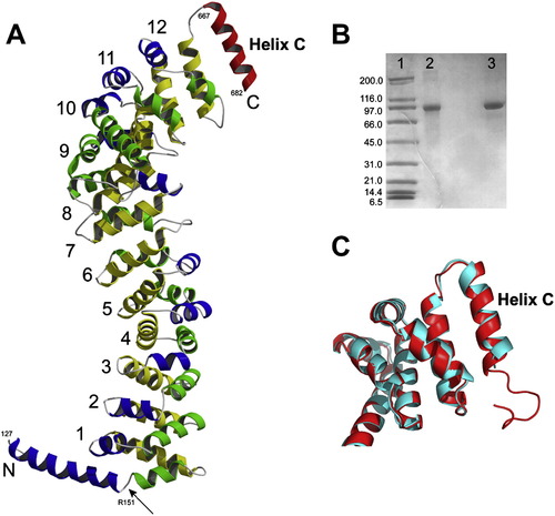
Full-Length Zebrafish β-Catenin Crystal Structure (A) Crystal structure of full-length zebrafish β-catenin. The color scheme of β-catenin armadillo repeats is the same as that in Figure 1. The C-terminal helix (helix C) is colored in red. The N-terminal helix is colored in blue and green with an arrow pointing to the kink. All β-catenin residue numbers shown in this figure are these of corresponding residue numbers of human β-catenin. (B) SDS-gel analysis of the dissolved full-length zebrafish β-catenin crystals. Lane 1, molecular weight marker. Lane 2, dissolved crystals. Lane 3, purified full-length zebrafish β-catenin. (C) Structural superposition between human β-catenin-R1C (red) and full-length zebrafish β-catenin (cyan) around helix C. |
Reprinted from Structure (London, England : 1993), 16(3), Xing, Y., Takemaru, K., Liu, J., Berndt, J.D., Zheng, J.J., Moon, R.T., and Xu, W., Crystal Structure of a Full-Length beta-Catenin, 478-487, Copyright (2008) with permission from Elsevier. Full text @ Structure