|
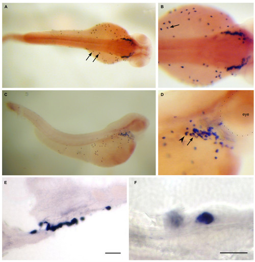
Dr-S100A11 expression pattern by whole mount in situ hybridization. Five day old zebrafish larvae were hybridized with RNA antisense probe. Panels A) to D), whole mounts; panels E) to F), sectioned after hybridization. A) Dorsal view, isolated large cells, mostly on the yolk sac, are labeled. B) Close up of A), an isolated cell with typical crescent-shaped soma signal is visible (arrow). C) Lateral view, a cluster of cells sits in the pericard, several cells are found on the yolk sac, a few in the skin. D) Close-up of the pericard region, expression intensity appears to vary (Arrowhead, arrow). E) Section through the cell cluster, scale bar 30 μm. F) Section with a single labeled cell, scale bar 10 μm.
|