|
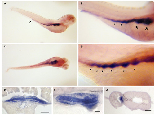
Dr-S100A10a expression pattern by whole mount in situ hybridization. Five day old zebrafish larvae were hybridized with RNA antisense probe. Panels A) to D), whole mounts; panels E) to F), sectioned after hybridization. Scale bars, 30 μm. A) Lateral view shows expression restricted to the whole intestinal tract including the anal region. B) Enlarged view, anterior is to the right, arrows point to the label in intestine. C) Ventral view, no other A10a-expressing regions are detected. D) Gut loops with high expression levels are pointed out by arrows. E, F) Cross sections of whole mount hybridizations at the level of the intestine. Only epithelial cells are strongly labeled, see white enclosure of a single epithelial cell in panel F). G) Gut epithelial cells are labeled as well.
|