
- Title
-
Structural and functional diversification in the teleost S100 family of calcium-binding proteins
- Authors
- Kraemer, A.M., Saraiva, L.R., and Korsching, S.I.
- Source
- Full text @ BMC Evol. Biol.
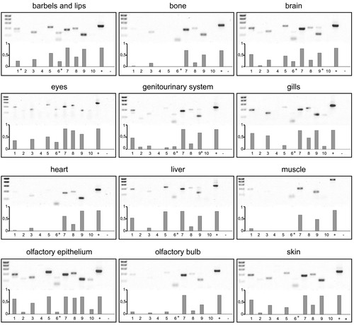
Expression of ten s100 genes in adult zebrafish by RT-PCR. mRNA was extracted from adult zebrafish tissue and transcribed into cDNA. Primer positions for PCR see Materials and Methods. Twelve different tissues were examined (barbels and lips, bone, brain, eyes, genitourinary tissue, gills, heart, liver, muscle, olfactory bulb, olfactory epithelium, skin). Band intensity was quantified to obtain an estimate of abundance to compare expression levels of several genes at least within the same tissue. Asterisks indicate bands at unexpected size (resulting from unspecific amplification) that were not quantified. EXPRESSION / LABELING:
|
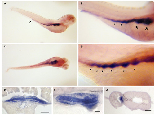
Dr-S100A10a expression pattern by whole mount in situ hybridization. Five day old zebrafish larvae were hybridized with RNA antisense probe. Panels A) to D), whole mounts; panels E) to F), sectioned after hybridization. Scale bars, 30 μm. A) Lateral view shows expression restricted to the whole intestinal tract including the anal region. B) Enlarged view, anterior is to the right, arrows point to the label in intestine. C) Ventral view, no other A10a-expressing regions are detected. D) Gut loops with high expression levels are pointed out by arrows. E, F) Cross sections of whole mount hybridizations at the level of the intestine. Only epithelial cells are strongly labeled, see white enclosure of a single epithelial cell in panel F). G) Gut epithelial cells are labeled as well. EXPRESSION / LABELING:
|

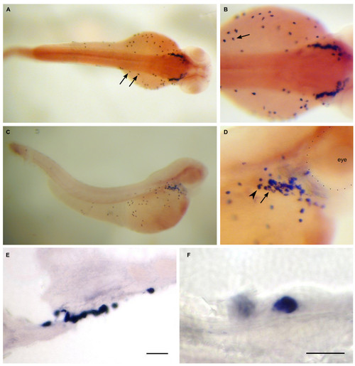
Dr-S100A10b expression pattern by whole mount in situ hybridization. Five day old zebrafish larvae were hybridized with RNA antisense probe. Panels A) to D), whole mounts; panels E) to F), sectioned after hybridization. Scale bars, 30 μm. A) Lateral view shows expression in the lower jaw. B) Lateral oblique view, lip region (arrows) expresses S100A10b. C) Frontal view (ventral to the right) shows expression in the mouth region. D) Ventral view (anterior is to the right), expression is visible in six branchial arches (asterisks), neuromasts (arrows) are not stained. E) Expression in branchial arches (asterisks) is limited to the epithelial layer. F) The seventh branchial arch (asterisk) is also expressing A10b, as well as cells of the pectoral fin (arrows). EXPRESSION / LABELING:
|
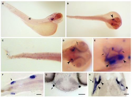
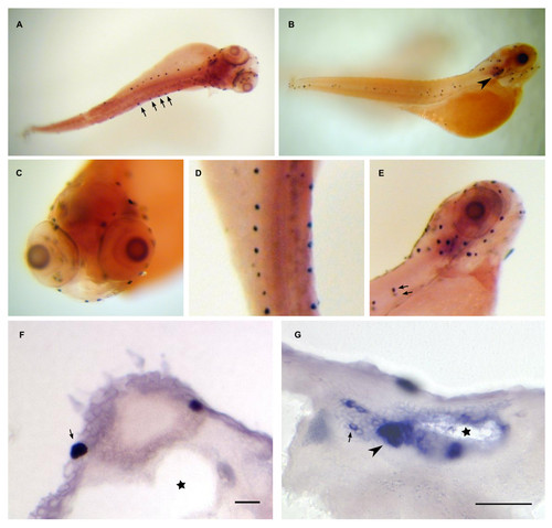
Dr-S100A11 expression pattern by whole mount in situ hybridization. Five day old zebrafish larvae were hybridized with RNA antisense probe. Panels A) to D), whole mounts; panels E) to F), sectioned after hybridization. A) Dorsal view, isolated large cells, mostly on the yolk sac, are labeled. B) Close up of A), an isolated cell with typical crescent-shaped soma signal is visible (arrow). C) Lateral view, a cluster of cells sits in the pericard, several cells are found on the yolk sac, a few in the skin. D) Close-up of the pericard region, expression intensity appears to vary (Arrowhead, arrow). E) Section through the cell cluster, scale bar 30 μm. F) Section with a single labeled cell, scale bar 10 μm. EXPRESSION / LABELING:
|

Dr-S100I.1 expression pattern by whole mount in situ hybridization. Five day old zebrafish larvae were hybridized with RNA antisense probe. Panels A) to G), whole mounts; panels H) to K), sectioned after hybridization. Scale bars 30 μm. A) Lateral view, strong ubiquitous expression is seen in the skin, the urogenital opening (bottom arrow), the rim of the dorsal fin (top row of arrows), the pectoral fin (arrowhead) and the lower jaw. B) Dorsal view, strong expression in the pectoral fin, the dorsal fin, and the lateral line (triangle-headed arrows) C) Enlargement of dorsal view, expression in the lateral line (arrows) and the dorsal fin (rim indicated by white spots). D) Larger magnification of urogenital opening (arrow) and the labeled lateral line (triangle-headed arrows) surrounding the neuromasts (diamond-headed arrows), which are not labeled. E) Ventral view, strong expression in branchial arches is seen. F) Enlargement of pectoral fin, especially the rim is heavily labeled. G) Dorsal view, expression in the olfactory placode (white dots). H) Cross section of the pectoral fin. I) Pharynx is intensely labeled. J) Three branchial arches (asterisks) are cross-sectioned; the mesenchyme including the cartilaginous bar is devoid of staining. K) The olfactory placode is heavily stained. EXPRESSION / LABELING:
|
Dr-S100S expression pattern by whole mount in situ hybridization. Five day old zebrafish larvae were hybridized with RNA antisense probe. Panels A) to E), whole mounts; panels F) to H), sectioned after hybridization. A) Lateral view, expression in neuromasts (arrows) and otic placode is visible. B) Dorsal view, staining in both otic placodes (arrowhead) is seen. C) Magnification, neuromasts in trunk and tail are labeled. D) Frontal view of the head region, expression in neuromasts (arrow) and the otic placode (arrowhead) can be seen. E) Magnification of the ear region, expression in several spots (arrowheads) in the otic placode and in climbing fibers (arrows). F) Expression in tail neuromasts, all hair cells seem to be stained (arrow), the ring of hair cell nuclei is devoid of staining. Scale bar 10 μm. G) Expression in a symmetrical pair of neuromasts in the head region, scale bar 30 μm. H) Staining in the ear, anterior is up. Hair cells underlying the otoliths show strong expression (arrowheads). A neuromast (arrow) is also visible on each side. Scale bar 30 μm. EXPRESSION / LABELING:
|
Dr-S100T expression pattern by whole mount in situ hybridization. Five day old zebrafish larvae were hybridized with RNA antisense probe. Panels A) to E), whole mounts; panels F) to G), sectioned after hybridization. Scale bars 30 μm. Dorsal (A) and lateral (B) view show expression in lateral line neuromasts (arrows) and the otic placode (arrowhead). C) Frontal view of the head region. Several labeled neuromasts are visible. D) Magnification of the trunk region from panel A), view from dorsal. E) Enlarged lateral view of the head region, over ten labeled neuromasts are seen. One neuromast and its contra lateral counterpart are indicated by arrows. EXPRESSION / LABELING:
|
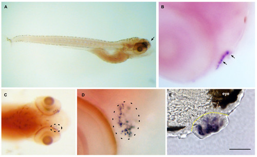
Dr-S100Z expression pattern by whole mount in situ hybridization. Five day old zebrafish larvae were hybridized with RNA antisense probe. Panels A) to D), whole mounts; panel E), sectioned after hybridization. Scale bar 30 μm. Lateral view. Expression in the larva is restricted to the olfactory placode (arrow). B) Closer view of the olfactory placode shows several large, labeled cells (arrows). C) Dorsal view of the head region, labeled cells in both olfactory placodes are visible, right placode encircled with dashed line. D) Enlarged view of a single olfactory placode (circled). Labeled cells are a subpopulation within the olfactory placode. E) Cross-section through an olfactory placode (delineated by yellow dashed line), several labeled cells are visible. EXPRESSION / LABELING:
|