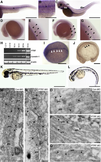
PTRF Is Required for Caveola Formation in Zebrafish Notochord Cells
(A–G) Expression pattern of ptrf during zebrafish development was analyzed by whole-mount mRNA in situ hybridization. Anterior is to the left and dorsal to the top. ptrf expression was detected in the notochord (n) in 24 hr embryos (A) and 31 hr embryos (B). In 72 hr embryos (C), expression was detected in dorsal blood vessels (dbv), in the otic vesicle (ov), and in the branchial arches (ba). ptrf mRNA expression was first detected in 18 hr embryos (F and G), and it was not detected in 16 hr embryos (D and E).
(H) RT-PCR comparing the temporal mRNA expression pattern of ptrf and cav1 during early embryo development. ptrf mRNA expression arose at 18 hr and was maintained throughout the development until 72 hr. cav1 mRNA expression was detected earlier than ptrf and was detected in the notochord as early as 12 hr by in situ hybridization (I). Expression of Cav1 protein was observed in the notochord by immunohistochemistry of 16 hr embryos (J). Arrowheads in (A)–(J) indicate the notochord.
(K and L) Ptrf knockdown embryos were generated by morpholino (MO) injection (6 ng/embryo). At 48 hr, 79% of control MO injected embryos appeared normal (K), whereas 44% of embryos injected with PTRF MO were curved under and/or presented heart edema (L).
(M–Q) Ultrastructural analysis and immunogold labeling of the notochord of 48 hr embryos injected with control MO (M and O) or PTRF MO (N, P, and Q). In the regions of cell-cell contact, the plasma membranes (PM) of neighboring cells are closely apposed. The plasma membrane in these regions is covered in caveolae (some of which are indicated by arrowheads) in the control MO-injected embryos (M). Caveolin immunogold labeling (arrowheads) is associated with the invaginated caveolae as seen in frozen sections labeled in parallel (O). The density of caveolae is greatly reduced in PTRF MO-injected embryos (N), and the plasma membranes of neighboring cells are less closely apposed. Caveolin labeling is associated predominantly with flat plasma membrane rather than invaginated caveolae in the PTRF MO-injected embryos (P and Q). pm, plasma membrane. Scale bars (A, B, C, D, F, I, J, K, and L), 250 μm, (E and G) 80 μm, and (M–Q) 200 nm.
|
