
Fig. 2
- ID
- ZDB-FIG-080303-6
- Publication
- Wen et al., 2008 - Visualization of monoaminergic neurons and neurotoxicity of MPTP in live transgenic zebrafish
- Other Figures
- All Figure Page
- Back to All Figure Page
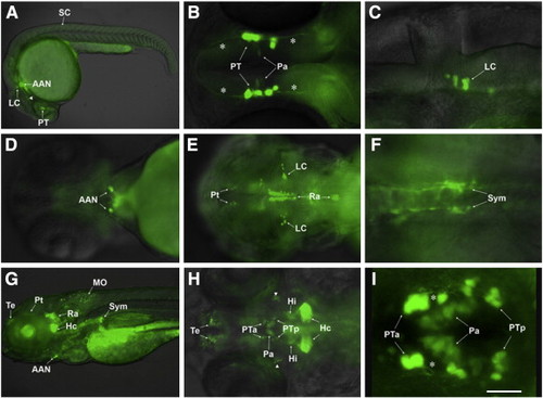
GFP expression pattern of ETvmat2:GFP embryos. Photos were taken under a fluorescence microscope in lateral (A, C, G), dorsal (B, E, F, H) and ventral views (D), or under a confocal microscope in dorsal view (I), rostral to the left. (A) A 24 hpf embryo showing the first GFP-positive (GFP+) neurons that appear in the nervous system; (B) a 30 hpf embryo showing GFP+ neurons in posterior tuberculum (PT) and their early axons projecting both anteriorly and posteriorly (asterisks), and some faintly labeled neurons in the paraventricular organ (Pa); (C) a 36 hpf embryo showing GFP+ neurons in the locus coeruleus (LC) located in the ventral lateral region of rhombomere 1; (D) a 54 hpf embryo showing the GFP+ arch-associated neurons (AAN) rostral to the heart; (E) a 60 hpf embryo with GFP+ neurons in the LC, the raphe nuclei (Ra) and the pretectum (Pt); (F) a 72 hpf embryo with GFP+ sympathetic neurons (Sym) under the artery; (G, H) 5 dpf embryos showing GFP+ neural groups in the central and peripheral nervous systems; (I) a 4 dpf embryo showing GFP+ neurons in PT and Pa, with the PT neurons sending out prominent single lateral neurites (asterisks). Scale bar: 25 μm. AAN: arch-associated neurons; Hc: caudal hypothalamus neural cluster; Hi: intermediate hypothalamus neural cluster; LC: locus coeruleus; MO: medulla oblongata neural cluster; Pa: neural cluster of paraventricular organ; Pt: pretectal neural cluster; PTa: anterior group of the posterior tubercular neurons; PTp: posterior group of the posterior tubercular neurons; Ra: raphe nuclei; SC: spinal cord; Sym: sympathetic neurons; Te: telencephalic neurons. Arrowheads in panels A and H: GFP-positive neurons in midbrain. |
| Gene: | |
|---|---|
| Fish: | |
| Anatomical Terms: | |
| Stage Range: | Prim-5 to Day 5 |
Reprinted from Developmental Biology, 314(1), Wen, L., Wei, W., Gu, W., Huang, P., Ren, X., Zhang, Z., Zhu, Z., Lin, S., and Zhang, B., Visualization of monoaminergic neurons and neurotoxicity of MPTP in live transgenic zebrafish, 84-92, Copyright (2008) with permission from Elsevier. Full text @ Dev. Biol.