|
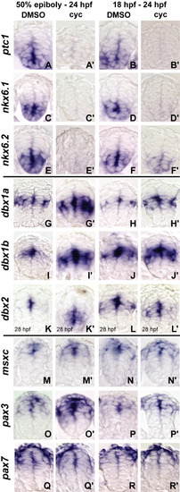
Analysis of patterning defects in cyclopamine-treated embryos. Spinal cord cross-sections of embryos treated with dimethyl sulfoxide (DMSO) or cyclopamine at 50% epiboly or 18 hours postfertilization (hpf) are shown. A-F: The 100 μM cyclopamine blocked expression of ptc1 (A-B″), nkx6.1 (C-D″), and nkx6.2 (E-F″) under both treatment conditions. G,I,K: Expression of dbx1a (G, G″), dbx1b (I, I″), and dbx2 (K, K″) expands ventrally after cyclopamine treatments beginning at 50% epiboly compared with DMSO controls. H,J,L: Similarly, after cyclopamine treatments beginning at 18 hpf, dbx1a (H,H″), dbx1b (J,J″), and dbx2 (L,L″) expand slightly ventrally. M-P: The expression of msxc (M-N″) is unaffected at either stage of treatment, whereas pax3 (O,O″) expression expands ventrally into the dbx1a/1b domain when treated at 50% epiboly, but no expansion is observed in the 18-24 hpf treatment (P,P″). Q,R: In contrast, pax7 (Q-R″) expression does not expand ventrally in either treatment.
|