
Fig. 8
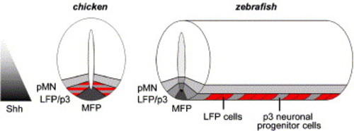
Schematic model of ventral neural tube organization during early neurogenesis in chicken and zebrafish. In chicken, non-neuronal LFP and p3 neuronal progenitor cell markers overlap in the domain between MFP and motoneuron progenitors (pMN). In zebrafish, LFP cells (in red) and p3 neuronal progenitor cells (in grey) alternate along the AP axis within this domain. During early embryonic development, distinct domains of cells that require different levels of Shh activity are generated along the DV axis of the vertebrate neural tube. In zebrafish, LFP and p3 neuronal progenitor cells, which are positioned at the same level along the DV axis, require different levels of HH activity. Note that MFP, LFP and neural progenitor domains are several cells wide in chicken, while in zebrafish MFP and LFP are only one cell in width and the pMN domain is two to three cells wide. |
Reprinted from Developmental Biology, 301(1), Schafer, M., Kinzel, D., and Winkler, C., Discontinuous organization and specification of the lateral floor plate in zebrafish, 117-129, Copyright (2007) with permission from Elsevier. Full text @ Dev. Biol.