
Fig. 1
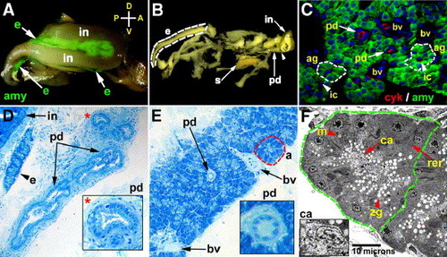
Adult exocrine pancreas. (A) Whole mount image of the dissected adult zebrafish digestive tract, right lateral view, processed for amylase immunohistochemistry (IHC). Exocrine tissue (green) is identified between adjacent intestinal loops. (B) Adult zebrafish pancreas dissected from adherent intestinal tissue. This fixed specimen reveals the branched network of exocrine lobules and associated ducts. Arrowhead points to insertion site of the extrapancreatic duct to the intestine. An exocrine lobule is outlined by the dashed white line. (C) Histological section of adult pancreas processed for cytokeratin (red) and amylase (green) IHC; DNA counterstained with dapi (blue) to visualize cell nuclei. Dashed white lines outline two acinar glands. Arrows point to large pancreatic ducts within the lobule. Arrowheads point to intercalated ducts. (D) Sagittal histological section showing large pancreatic ducts with adjacent exocrine and intestinal tissues. (E) Histological section of an exocrine lobule showing acinar cells with a prominent duct. Insets (* in panel D; long arrow in panel E) show cuboidal cells lining these ducts. Red dashed line outlines an acinus. (F) Transmission electron micrograph showing an acinus comprised of polarized cells with apical zymogen granules, basal nucleus, mitochondria and rough endoplasmic reticulum. A centroacinar cell is identified based upon its position within the acinar lumen and its scant cytoplasm (inset). e: exocrine tissue; in: intestine; amy: amylase; s: spleen; ag: acinar glands; pd: pancreatic ducts; ic: intercalated ducts; bv: blood vessel; a: acinus; zg: zymogen granules; m: mitochondria; rer: rough endoplasmic reticulum; ca: centroacinar cell; A: anterior; P: posterior; D: dorsal; V: ventral. |
Reprinted from Developmental Biology, 284(1), Yee, N.S., Lorent, K., and Pack, M., Exocrine pancreas development in zebrafish, 84-101, Copyright (2005) with permission from Elsevier. Full text @ Dev. Biol.