
Fig. 7
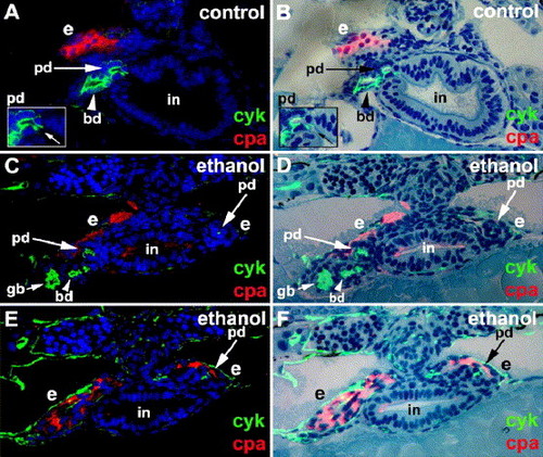
Ethanol treatment disrupts exocrine pancreas morphogenesis. Histological cross-sections (viewing posteriorly) of a 4 dpf control (A, B) and an ethanol-treated larva (C–F) processed for cyk (green) and cpa (red) immunohistochemistry. (A, C, E) Fluorescent images with dapi (blue); (B, D, F) merged fluorescent images without dapi and bright field images of the corresponding histological sections stained with methylene blue and azure II. (A, B) The extrapancreatic duct and extra-hepatic bile duct of control larvae join the intestine at the same level. Inset shows site of pancreatic duct insertion into the intestine (arrow). (C–F) Bilateral cyk+ and cpa+ exocrine tissues are present in ethanol-treated larva. Bilateral extrapancreatic ducts that fail to join the intestine in these sections, as well as in consecutive rostral and caudal sections (not shown). Sections depicted in panels (C), (D) and (E), (F) are separated by 9 μm (C and D rostral to E and F). cyk: cytokeratin; cpa: carboxypeptidase A; pd: pancreatic duct; bd: bile duct; in: intestine; e: exocrine tissues; gb: gall bladder. |
| Gene: | |
|---|---|
| Fish: | |
| Condition: | |
| Anatomical Term: | |
| Stage: | Day 4 |
Reprinted from Developmental Biology, 284(1), Yee, N.S., Lorent, K., and Pack, M., Exocrine pancreas development in zebrafish, 84-101, Copyright (2005) with permission from Elsevier. Full text @ Dev. Biol.