
Fig. 4
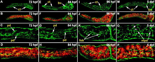
Pancreatic ductal morphogenesis during larval development. (A–P) Confocal images (right, lateral view) of whole mount specimens processed for cyk (green) and cpa (red) immunohistochemistry. (A–D) At 72 hpf, small primary pancreatic ducts are evident, and there is strong cpa staining in acinar cells. (E–H) The 84 hpf pancreas has a few more ducts than at earlier stages. (I–L) At 96 hpf, first order ductal branches are evident. These small ducts appear to extend into the nearby acinar glands. (M–P) At 120 hpf (5 dpf), second order ductal branches (double arrows) that clearly extend into well formed acini are evident. First order ductal branches (single arrow) and main pancreatic duct segments are also evident at this stage. cyk: cytokeratin; cpa: carboxypeptidase A; pd: pancreatic ducts; fob: first order ductal branches; mpd: main pancreatic duct; ct: connective tissue; bv: blood vessel. |
| Gene: | |
|---|---|
| Fish: | |
| Anatomical Term: | |
| Stage Range: | Protruding-mouth to Day 5 |
Reprinted from Developmental Biology, 284(1), Yee, N.S., Lorent, K., and Pack, M., Exocrine pancreas development in zebrafish, 84-101, Copyright (2005) with permission from Elsevier. Full text @ Dev. Biol.