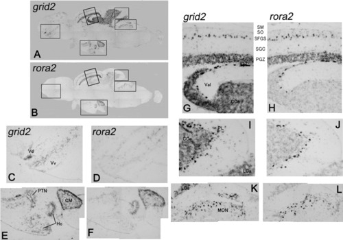
Expression pattern of grid2, glutamate receptor delta2 (A,C,E,G,I,K) and rora2 (B,D,F,H,J,L) was detected by in situ hybridization in adjacent sections of an adult brain of zebrafish. High magnification views of the boxed regions in A and B are shown in the ventral part of the telencephalon (C,D), the ventral part of the midbrain (E,F), the optic tectum and anterior part of the cerebellum (G,H), the posterior part of the cerebellum (I,J), and the dorsal part of the pons (K,L). Colocalized expression of these two genes was observed in the cerebellar-like structures. Abbreviations are the same as used in Wullimann et al. ([1996]). Vdm, dorsal nucleus of V; Vv, ventral nucleus of V; PTN, posterior tuberal nucleus; Hc, caudal zone of periventricular hypothalamus; CM, corpus mamillare; SM, stratum marginale; SO, stratum opticum; SFGS, Stratum fibrosum et griseum superficiale; SGC, stratum griseum centrale; PGZ, periventricular gray zone; Val, lateral division of valvula cerebelli; CCe, corpus cerebelli; PL, Purkinje cell layer; LCa, lobus caudalis cerebelli; MON, medial octavolateralis nucleus.
|
