FIGURE

Fig. 3

ID
ZDB-FIG-070911-92
Publication
Katsuyama et al., 2007 - Expression of zebrafish ROR alpha gene in cerebellar-like structures
Other Figures
All Figure Page
Back to All Figure Page
Fig. 3

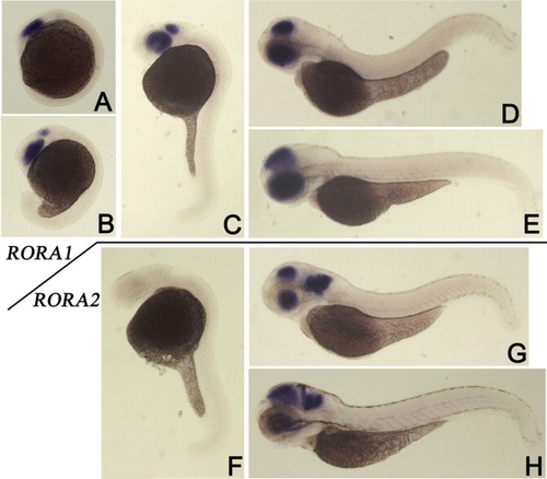
A-H: Lateral views of in situ hybridization specimens in which expression of rora1 (A-E) or rora2 (F-H) was detected. A: At the five-somite stage (12 hours postfertilization [hpf]). B: At the 18-somite stage (18 hpf). C,F: Expression of rora1 (C) was observed in the eyes and midbrain, but expression of rora2 (F) was not detected in 24 hpf specimens. D,G: Expression of rora1 (D) and rora2 (G) was similarly observed in the eyes and tectum, but only rora2 expression was observed in the hindbrain of 2 days postfertilization (dpf) specimens (G). H: Expression of rora2 in the cerebellum is observed in 3 dpf specimens.

Expression Data
Genes:
Fish:
Anatomical Terms:
Stage Range: 5-9 somites to Protruding-mouth

Expression Detail
Antibody Labeling
Phenotype Data

Phenotype Detail
Acknowledgments
This image is the copyrighted work of the attributed author or publisher, and ZFIN has permission only to display this image to its users. Additional permissions should be obtained from the applicable author or publisher of the image. Full text @ Dev. Dyn.