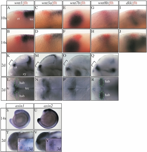
Expression of various Wnt pathway genes in and around the epithalamus. (A-K,M,O,Q,S-V) Lateral views and (L,N,P,R,T′,V′) dorsal views focused on the diencephalic epithalamus (et; except S,U). All developmental stages are indicated on the left of each horizontal row of panels and the marker genes used on the top of each vertical row. (A-J) floating head (flh) expression in the epithalamus (in red) has been used as a landmark. (I,J) dickkopf (dkk) is expressed in the epithalamic area at mid-somitogenesis stages. Its expression fades shortly later (data not shown). (S,T,T′) In zebrafish and Xenopus axin1 is initially expressed ubiquitously up to tailbud stages but becomes subsequently confined to the fore- and midbrain region (Hedgepeth et al., 1999; Van de Water et al., 2001). (T′) Similar to Xenopus we find increased axin1 expression in the habenula from three days of development onward, but more pronounced on the left side (red dotted circle). (U,V,V′) axin2 is expressed in the dorsal neural tube and CNS including the epithalamus also at later stages. For further details see supplementary results. ey, eye; hab, habenula (green circle); tc, telencephalon; tec, tectum.
|
