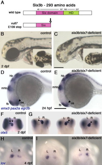
Combined Loss of Six3b/Six7 Function Results in Lack of Eye Tissue and Abnormal Brain Laterality (A) Schematic presentation of normal Six3b and the predicted truncated Six3b protein, encoded by the six3bvu87 allele. (B and C) Live control embryo (B) and a six3b/six7-deficient embryo (B) demonstrating lack of eye tissue. (D and E) Comparable brain patterning in 24 hpf control (D) and six3b/six7-deficient (E) embryos. Forebrain (fb), midbrain-hindbrain boundary (mhb), and hindbrain (hb) are labeled with emx3, pax2a, and egr2b, respectively. The optic stalk (os, arrowheads, pax2a) is reduced in the six3b/six7-deficient embryo. (F and G) Pineal (arrow) and parapineal localization (arrowheads) in control (F) and six3b/six7-deficient (G) embryos. (H and I) Habenular nuclei labeled with lov, in control (H) and six3b/six7-deficient (I) embryos. Control embryos are six3bvu87/+ or six3bvu87/vu87 and present normal phenotypes. R, right; L, left. Anterior is to the left in (B)–(E) and up in (F)–(I). (B)–(E) are lateral views, and (F)–(I) and insets in (B) and (C) are dorsal views. Scale bars, 200 μm.
|
