|
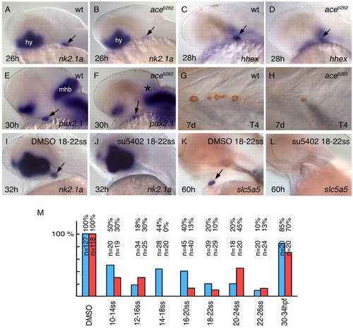
FGF signalling is required for thyroid development. Labelling is as in Fig. 1. Anterior is to the left. Lateral (A-F,I-L) and ventral (G,H) views are shown. (A-F) Expression of thyroid markers in ace mutants and in wild-type (wt) siblings. (G,H) ace larvae have a strong reduction in thyroid gland size. (I,K) In DMSO-treated control embryos, the thyroid appears normal. (J,L,M) Following different stages of su5402 treatment, the thyroid is completely lost. (J,L) Example embryos without thyroid; (M) the complete data set. Blue bars, embryos with thyroidal nk2.1a expression; red bars, slc5a5 expression. Arrows point to the thyroid primordium; the asterisk indicates the absent midbrain-hindbrain boundary (MHB) in ace mutants.hy, hypothalamus; mhb, midbrain-hindbrain boundary, ss somite stage.
|