
Fig. 4
- ID
- ZDB-FIG-070813-20
- Publication
- Wendl et al., 2007 - Early developmental specification of the thyroid gland depends on han-expressing surrounding tissue and on FGF signals
- Other Figures
- All Figure Page
- Back to All Figure Page
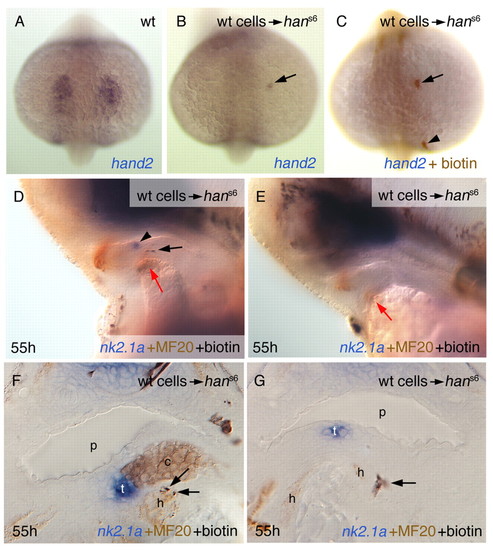
Grafted wild-type cells can restore nk2.1a expression in hans6 mutant embryos. Labelling of panels is as in Fig. 1. Anterior up (A-C) or to the left (D,E), and sections (F,G). (A-C) Grafted wild-type cells express han (hand2) in the mutant background. Arrows point to a single grafted cell that contributed to the anterior lateral plate mesoderm. The arrowhead in C indicates a grafted cell that contributed to other tissue, not expressing hand2. (D-E) Examples of hosts fixed at 55 hours post fertilisation (hpf). D shows an embryo (corresponding to #4 in Table 1) that has restored nk2.1a expression (arrowhead), an occurrence that was never seen in siblings. The black arrow points to grafted wild-type cells, the red arrows to the heart rudiment. E shows a hans6 embryo without restored thyroidal nk2.1a expression. (F,G) Cross sections of specimens of hans6 embryos with restored nk2.1a expression. Grafted cells in the embryo shown in F (corresponding to #5 in Table 1) are adjacent to the thyroid in cartilage, but also in heart tissue (arrows). Note also that such a large thyroid as in F was never observed in untreated hans6 embryos. Often, donor cells clearly belong to the pharyngeal mesenchyme, but are also close to the heart rudiment (G, embryo corresponds to #3 in Table 1). c, cartilage precursor cells; h, heart rudiment; p, pharynx; t, thyroid. |