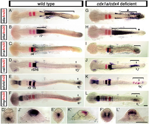
Loss of Cdx function activates the expression of hindbrain genes in the posterior CNS. Expression of hindbrain markers (purple) in wild-type (A-F,D'-F') and cdx1a/cdx4-deficient (G-L,J'-L') zebrafish embryos counterstained for the r3 and r5 marker krx20 (red signal). (A,G) Spinal cord hoxb8a expression (bracket in A) is lost in cdx1a/cdx4-deficient embryos (bracket with asterisk in G). (B,H) r7/8 hoxd4a expression (bracket in B) is expanded caudally in cdx1a/cdx4-deficient embryos (bracket in H) except for the most caudal tip of the CNS (asterisk in H). (C,I) In addition to its normal domain of expression in r4, hoxb1a expression can also be seen in the posterior CNS of cdx1a/cdx4-deficient embryos (bracket in I). (D,J) In the hindbrain, val is expressed in r5 and r6 of wild-type (D) and cdx1a/cdx4-deficient (J) embryos. In the tail region, val is also expressed in the posterior CNS of cdx1a/cdx4-deficient (bracket in J,J') but not wild-type (D') embryos. (E,K) Overlapping expression of epha4a (purple) and krx20 (red) are visualized in r3 and r5 of wild-type (E) and cdx1a/cdx4-deficient (K) embryos. In the tail region, epha4a and krx20 are expressed in the posterior CNS of cdx1a/cdx4-deficient (bracket in K,K') but not wild-type (E') embryos. (F,L) In the hindbrain, radical fringe (rfng) is expressed in seven stripes at the rhombomere boundaries in wild-type (F) and cdx1a/cdx4-deficient embryos (L). In the tail region, rfng is also expressed in the posterior CNS of cdx1a/cdx4-deficient (bracket in L,L') but not wild-type (F') embryos. For each condition, a minimum of 44 embryos from at least three independent experiments were analyzed, with more than 82% of embryos displaying the phenotype shown. Representative 20-somite, stage-matched, whole-mounted embryos are shown in dorsal view, anterior to the left. The position of somite 3, the hindbrain-spinal cord transition in wild-type embryos, is indicated with a white arrowhead. The planes of section are indicated with two short vertical bars. Sections are dorsal to the top, with the neural rod delineated by the dashed line. Scale bars: 100 µm.
|
