
Fig. 3
- ID
- ZDB-FIG-070416-3
- Publication
- Krock et al., 2007 - Noncell-autonomous photoreceptor degeneration in a zebrafish model of choroideremia
- Other Figures
- All Figure Page
- Back to All Figure Page
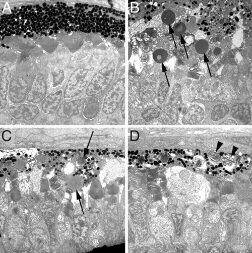
Transmission electron microscopy of 4.5 dpf wild-type and rep1 mutant retinas. (A) Electron micrographs of wild-type retinas reveal an orderly array of photoreceptor outer segments and the uniform thickness of the RPE. Melanosome maturation within the RPE is normal. (B–D) Electron micrographs from multiple rep1 retinas showing degeneration of the RPE and photoreceptors. Arrows in B and C indicate large vacuoles observed in the RPE of rep1 mutants. Arrowheads in D indicate outer segment material not digested by the RPE. Melanosome size and maturation vary more dramatically and are less dense than what is seen in wild type. |
| Fish: | |
|---|---|
| Observed In: | |
| Stage: | Day 4 |