
Fig. 2
- ID
- ZDB-FIG-070416-2
- Publication
- Krock et al., 2007 - Noncell-autonomous photoreceptor degeneration in a zebrafish model of choroideremia
- Other Figures
- All Figure Page
- Back to All Figure Page
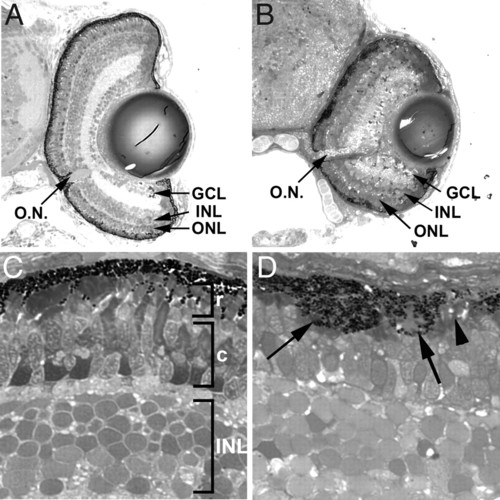
Histological sections of 4.5 dpf wild-type and rep1 mutant retinas. (A) Wild-type retinas at 4.5 dpf have fully laminated and retinal ganglion cells (GCL), amacrine and bipolar interneurons (INL), and photoreceptors (ONL) differentiated, and the optic nerve (O.N.) is apparent. (B) Lamination and cellular differentiation is not affected in rep1 mutants, but eye size is reduced and the RPE layer appears irregular. (C) High magnification of sections from light-adapted wild-type retinas showing the rod (r) outer segments positioned distally from the cone (c) outer segments. (D) Sections of rep1 mutants showing areas of RPE hypertrophy (arrows) into the photoreceptor layer and other regions where the RPE is almost devoid of pigmentation (arrowhead). Rod and cone outer segments are not normally positioned and are shorter than wild-type. |
| Fish: | |
|---|---|
| Observed In: | |
| Stage: | Day 4 |