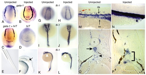
Analysis of gene expression during hematopoiesis and the formation of the trunk vasculature. (A-D) 8- to 10-somite stage embryos; posterior views with dorsal to the top. (A,B) Expression of gata1 in uninjected (A) and hrtMO(1)-injected (B) embryos. Note that injected embryos express gata1 in the most posterior region of the embryo, in contrast to uninjected embryos. (C) Expression of hrT and gata1 in uninjected embryo. (D) Expression of hrT in hrTMO(1)-injected embryo. The same expression pattern is found in uninjected embryos (not shown). Note that the hrT expression domain coincides with the region of ectopic gata1 expression in the morphants (B). (E,F) Live pictures of the posterior half of uninjected (E) and hrTMO(1)-injected (F) embryos at 36 hpf. Arrow indicates the blood pooling in the peri-anal region of the injected embryos. (G,H) Posterior views with dorsal towards the top. fli1 is expressed in a `U′-shaped pattern at 14 hpf in uninjected (G) and hrTMO(1)-injected (H) embryos. (I,J) Dorsal views with anterior towards the top. Expression of fli1 at 20 hpf. In uninjected embryos, fli1-expressing cells are at the midline (I), whereas fli1-expressing cells have not converged completely to the midline in hrT morphants (J). (K,L) Lateral views with anterior towards the top. The expression pattern of fli1 in uninjected (K) and hrTMO(1)-injected embryos at 24 hpf. White arrowhead indicates the fli1 expression in the pharyngeal primordium. (M,N) Lateral views of 24 hpf embryos with anterior towards the left. In uninjected embryos (M), fli1 expression is apparent in the dorsal aorta (da), axial vein (av) and intersegmental vessels (is), whereas hrT morphants (N) exhibit a single domain of fli1 expression in the midline of the entire trunk with no intersegmental vessels. (O,P) Transverse sections of embryos at 30 hpf stained with fli1, dorsal towards the top. In uninjected embryos (O), the formation of the dorsal aorta (black arrow) below the notochord (n) and axial vein (black arrowhead) is apparent. In the hrT morphants (P), only a single lumen is present. The black bracket indicates the developing region of the dorsal aorta and axial vein. Injected embryos were injected with 1.5 ng of hrTMO(1).
|
