
Fig. 3
- ID
- ZDB-FIG-070206-4
- Publication
- Rojas et al., 2007 - Cloning of hif-1alpha and hif-2alpha and mRNA expression pattern during development in zebrafish
- Other Figures
- All Figure Page
- Back to All Figure Page
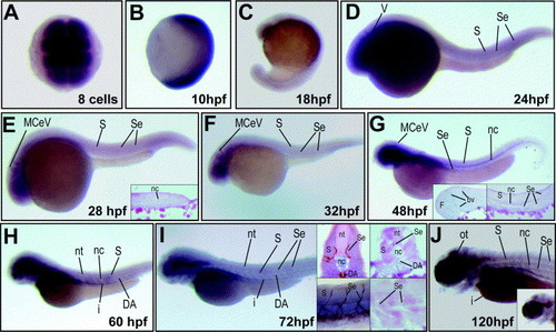
Expression of hif-2α in zebrafish embryos and larvae as revealed by mRNA in situ hybridization. hif-2α mRNA is maternally expressed and ubiquitously distributed at early stages (A and B), becoming ventrally restricted later in embryogenesis (C); (A) dorsal view; (B) and (C), lateral views with the anterior side on top. At 24 (D), 28 (E) and 32 hpf (F) hif-2α was detected in the brain, somites, and blood vessels. At 48 hpf, hif-2α expression is present in brain blood vessels, notochord and intersegmental blood vessels (G), as it is shown in the brain (left inset in G) and tail paraffin cross-sections (right inset in G). At 60 hpf, hif-2α mRNA is expressed in the brain, somites, dorsal aorta, intestine and notochord (H). At 72 hpf, hif-2α expression was detected in the brain, brain blood vessels, intersegmental vessels, dorsal aorta and intestine (I). mRNA in situ hybridization for hif-2α and anti-GFP immunohistochemistry in Tg(fli1:EGFP)y1 embryos show that expression occurs in intersegmental blood vessels (left-top inset in I, transverse cross-section; left-bottom inset, DIC lateral view). Paraffin cross-sections of wild type embryos at 72 hpf, show expression of hif-2α in somites, intersegmental blood vessels, notochord and aorta (right-top inset in I, transverse cross-section; right-bottom in I, sagittal cross-section). At 120 hpf, expression of hif-2α was detected in the optic tectum, intersegmental blood vessels, notochord, intestine and retina (J). (D)–(J) are lateral views with anterior to the left. Insets show transverse cross-sections with the dorsal part on the top, or sagittal cross-sections with anterior to the left. Abbreviations: V, ventricle epithelia; F, forebrain; M, midbrain; H, hindbrain; MCeV, middle cerebral vein; Se, intersegmental vessel; DA, dorsal aorta; nc, notochord; S, somites; nt, neural tube; I, intestine; bv, blood vessel; nm, neuromasts; ot, optic tectum; e, eye; r, retina. |
| Gene: | |
|---|---|
| Fish: | |
| Anatomical Terms: | |
| Stage Range: | 8-cell to Day 5 |
Reprinted from Gene expression patterns : GEP, 7(3), Rojas, D.A., Perez-Munizaga, D.A., Centanin, L., Antonelli, M., Wappner, P., Allende, M.L., and Reyes, A.E., Cloning of hif-1alpha and hif-2alpha and mRNA expression pattern during development in zebrafish, 339-345, Copyright (2007) with permission from Elsevier. Full text @ Gene Expr. Patterns