
Fig. 2
- ID
- ZDB-FIG-070206-1
- Publication
- Rojas et al., 2007 - Cloning of hif-1alpha and hif-2alpha and mRNA expression pattern during development in zebrafish
- Other Figures
- All Figure Page
- Back to All Figure Page
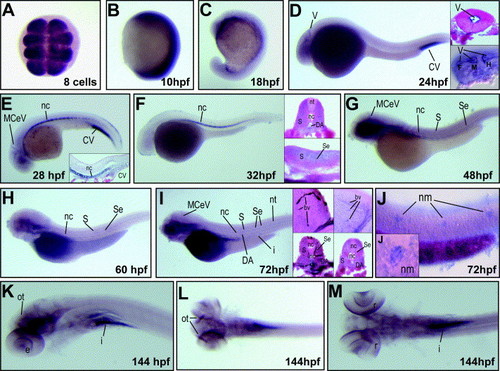
Whole-mount in situ hybridization for hif-1α. Expression of hif-1α mRNA was analyzed at the indicated developmental stages. hif-1α mRNA is detected at early stages in an ubiquitous expression pattern (A and B), becoming ventrally restricted by late embryogenesis (C); A, dorsal view; B and C, lateral view with anterior on top. At 24 hpf hif-1α is expressed in ventricle epithelia (D, paraffin transverse cross-section in top inset), as confirmed by Nomarski microscopy (D, bottom inset). At 28 hpf, expression is present in the notochord, caudal vein and brain blood vessels (E), as confirmed by histological sagittal cross-section (inset in E). At 32 hpf, hif-1α mRNA is detected in brain and notochord (F), paraffin cross-section of embryos shows that expression occurs in the somites, notochord and dorsal aorta (transverse cross-section, top inset in F) as well as in intersegmental blood vessels (sagittal cross-section, bottom inset in F). At 48 and 60 hpf (G and H, respectively) the expression of hif-1α is detected in the brain, notochord, somites and intersegmental blood vessels. At 72 hpf the mRNA is detected in the brain, branchial region, intersegmental blood vessels, dorsal aorta and intestine (I), left insets in I show a Tg (fli1:EGFP)y1 embryo subjected to GFP immunohistochemistry and hif-1α mRNA in situ hybridization; right insets in I show the mRNA in situ hybridization pattern of hif-1α in a wild type fish (all insets are transverse cross-sections). Overstaining of 72 hpf embryos revealed expression in caudal neuromasts (J), as confirmed by DIC optics (inset in J). At 144 hpf expression was observed in the optic tectum, retina and intestine (K). A dorsal view of embryos at 144 hpf shows expression in the optic tectum (L) and retina (M). In (D) to (M) lateral view, with anterior to the left. Insets showing transverse cross-sections with dorsal side up; sagittal cross-sections show the anterior to the left. Abbreviations: V, ventricle epithelia; F, forebrain; M, midbrain; H, hindbrain; MCeV, middle cerebral vein; CV, caudal vein; Se, intersegmental vessel; DA, dorsal aorta; nc, notochord; S, somites; nt, neural tube; I, intestine; bv, blood vessel; nm, neuromasts; ot, optic tectum; e, eye; r, retina. |
| Gene: | |
|---|---|
| Fish: | |
| Anatomical Terms: | |
| Stage Range: | 8-cell to Day 6 |
Reprinted from Gene expression patterns : GEP, 7(3), Rojas, D.A., Perez-Munizaga, D.A., Centanin, L., Antonelli, M., Wappner, P., Allende, M.L., and Reyes, A.E., Cloning of hif-1alpha and hif-2alpha and mRNA expression pattern during development in zebrafish, 339-345, Copyright (2007) with permission from Elsevier. Full text @ Gene Expr. Patterns