
Fig. 1
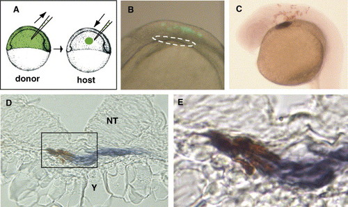
Neural crest cells migrate to the primary heart field and express cmlc2. (A) Transplantation strategy. At the shield stage, 10–50 prospective neural crest cells were transplanted from fluorescein dextran donors into the orthotopic region of hosts. (B) 8 SS embryo, DIC and epi-fluorescent (lateral view, anterior at left, dorsal at top). Lineage-labeled cells (green) were in neural crest and neural tube at hindbrain level, but not in anterior LPM (white circle). (C) 26 SS embryo, lineage-labeled cells (anti-fluorescein antibody, brown) and cardiomyocytes (cmlc2, purple) were detected. (D) Transverse section through the primary heart field of 26 SS embryo with lineage-labeled neural crest cells (brown) expressing cmlc2 (purple). NT = neural tube, Y = yolk. (E) Magnified image of box in panel D. |
| Gene: | |
|---|---|
| Fish: | |
| Anatomical Term: | |
| Stage: | 26+ somites |
Reprinted from Developmental Biology, 298(1), Sato, M., Tsai, H.J., and Yost, H.J., Semaphorin3D regulates invasion of cardiac neural crest cells into the primary heart field, 12-21, Copyright (2006) with permission from Elsevier. Full text @ Dev. Biol.