
Fig. 5
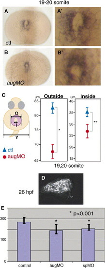
sema3D morphants have fewer cardiomyocytes in the primary heart field. Dorsal views cmlc2 expression in control (A, A′) and sema3D morphants (B, B′). (C) Schematic diagram of inside (I) and outside (O) diameters of cmlc2 expression (purple) and (Y) yolk diameter. Graphs indicate mean ± one standard deviation of diameters measured (μm) in sema3D morphants (red circles, n = 18) and controls (blue triangles, n = 13). The primary heart field is significantly smaller in sema3D morphants (*P = 0.0002, **P = 0.0549). (D) An image of dissociated cmlc2-gfp-positive heart at the 26 hpf, after arrival on neural crest. (E) Count of number of cardiomyocytes from dissociated cmlc2::EGFP embryos. y axis is mean number of EGFP-positive cardiomyocytes, bars indicate standard deviation. Control embryos had 184 ± 15 (n = 30) cardiomyocytes. Both aug-MO and sp-MO-injected embryos had significantly fewer GFP-positive cardiomyocytes (aug-MO: 145 ± 21, n = 23, sp-MO: 154 ± 11, n = 11) than control (*P < 0.0001). |
| Gene: | |
|---|---|
| Fish: | |
| Knockdown Reagent: | |
| Anatomical Term: | |
| Stage: | 14-19 somites |
Reprinted from Developmental Biology, 298(1), Sato, M., Tsai, H.J., and Yost, H.J., Semaphorin3D regulates invasion of cardiac neural crest cells into the primary heart field, 12-21, Copyright (2006) with permission from Elsevier. Full text @ Dev. Biol.