
Fig. S11
- ID
- ZDB-FIG-060914-1
- Publication
- Lin et al., 2006 - PPM1A functions as a Smad phosphatase to terminate TGFbeta signaling
- Other Figures
- All Figure Page
- Back to All Figure Page
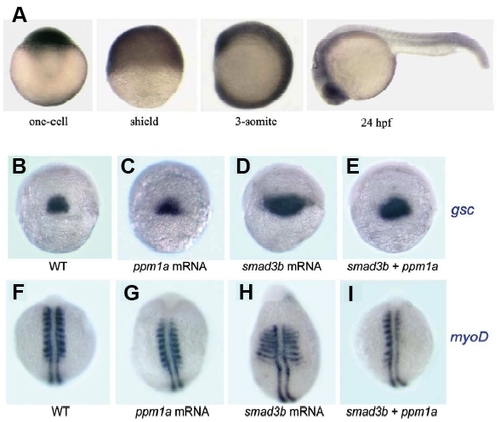
Expression Pattern and Smad3-Antagonizing Activity of PPM1A (A) Spatiotemporal expression pattern of PPM1A at different stages, detected by whole-mount in situ hybridization. Note that PPM1A transcripts are present ubiquitously. (B-I) Dorsal views showing gsc (B-E) expression at the shield stage and and myoD (E-I) at the 10-somite stage. mRNA species used for injection were indicated below each picture. WT, uninjected wildtype embryo. Injection with 50 pg Smad3b mRNA caused expansion of gsc expression at the shield stage in 63.4% (n=41) of embryos (D) and ventral expansion of the myogenic marker myoD in somites at the 10-somite stage in 64.5% (n=62) of embryos (H), indicating a dorsalizing effect of Smad3b. When the same amount of Smad3b mRNA and 300 pg PPM1A mRNA were coinjected, embryos with expanded gsc and myoD expression decreased to 30% (n=40) and 11.3% (n=71), respectively (E, I). These data indicate that effect of Smad3b expression can be inhibited by excess PPM1A in vivo. |
| Genes: | |
|---|---|
| Fish: | |
| Anatomical Terms: | |
| Stage Range: | 1-cell to Prim-5 |
Reprinted from Cell, 125(5), Lin, X., Duan, X., Liang, Y.Y., Su, Y., Wrighton, K.H., Long, J., Hu, M., Davis, C.M., Wang, J., Brunicardi, F.C., Shi, Y., Chen, Y.G., Meng, A., and Feng, X.H., PPM1A functions as a Smad phosphatase to terminate TGFbeta signaling, 915-928, Copyright (2006) with permission from Elsevier. Full text @ Cell