
Fig. 6
- ID
- ZDB-FIG-070919-21
- Publication
- Lin et al., 2006 - PPM1A functions as a Smad phosphatase to terminate TGFbeta signaling
- Other Figures
- All Figure Page
- Back to All Figure Page
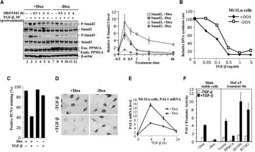
Inducible Expression of PPM1A Causes TGFβ Resistance(A) Reduced Smad2/3 phosphorylation in Mv1Lu cells with inducible expression of PPM1A. Mv1Lu-tet-off cells stably harboring Flag-PPM1A were grown with (+) or without (−) 10 ng/ml of doxycycline (Dox). Left: Induced expression of Flag-PPM1A (lanes 7–12). Cells were pretreated with TGFβ for 30 min (lanes 2 and 8) and then SB431542 for the indicated time periods (lanes 3–6 and 9–12). Levels of P-Smads, total Smads, and PPM1A were detected by Western blotting. Right: Line graph representing data from two stable clones with two experiments each, with values and error bars representing mean and standard deviation. (B) PPM1A causes partial TGFβ resistance. Mv1Lu-PPM1A cells were cultured ±Dox, treated with various doses of TGFβ, and subjected to [3H]thymidine incorporation to analyze DNA synthesis. (C) PPM1A blocks the TGFβ antiproliferative response. Mv1Lu-PPM1A cells, cultured ±Dox and treated ±TGFβ (0.2 ng/ml, 12 hr), were stained for PCNA (Zymed). PCNA-positive (black) and -negative (light gray) cells were counted and plotted. (D) A representative field of PCNA staining as in (C). (E) qRT-PCR analysis of PAI-1 in Mv1Lu cells. Values and error bars represent the mean and standard deviation of two experiments. (F) PPM1A inhibits PAI-1 promoter activity. Left: Stable Mv1Lu cells were transfected with p800-luc and grown with (+) or without (−) Dox. Right: HaCaT cells were transiently transfected with p800-luc and PPM1A expression plasmids. Values and error bars represent the mean and standard deviation of at least three experiments. |
Reprinted from Cell, 125(5), Lin, X., Duan, X., Liang, Y.Y., Su, Y., Wrighton, K.H., Long, J., Hu, M., Davis, C.M., Wang, J., Brunicardi, F.C., Shi, Y., Chen, Y.G., Meng, A., and Feng, X.H., PPM1A functions as a Smad phosphatase to terminate TGFbeta signaling, 915-928, Copyright (2006) with permission from Elsevier. Full text @ Cell