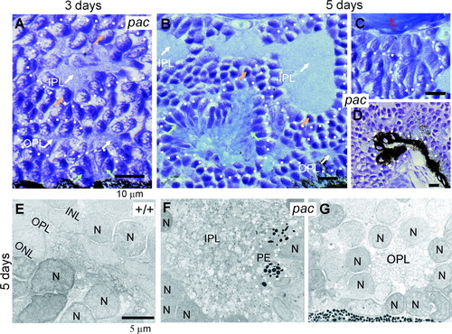
Rosette formation in the pac mutant. Arrows are as in Figure 1. A: Larger magnification of Figure 1D, showing three types of retinal rosettes at the 3-day stage: rosettes containing islands of inner plexiform layer tissue (IPL), rosettes containing islands of outer plexiform layer tissue (OPL), and rosettes of photoreceptors (green arrows). B: Larger magnification of Figure 1J, showing the same types of rosettes as in A at the 5-day stage. C: Cell cluster or rosette formed by retinal ganglion cells at day 5, magnified from Figure 1J. D: Rosette-like structures of the pigment epithelium, larger magnification of Figure 1L. E-G: Electron photomicrographs of 5-day-old retinae. E: WT outer plexiform layer and surrounding tissue. INL, inner nuclear layer; ONL, outer nuclear layer containing photoreceptor nuclei; N, nucleus. F: Mutant inner plexiform layer island and surrounding nuclei of the inner nuclear layer. Some mutant retinae contain small pigment epithelium (PE) islands within other retinal tissue. G: Mutant outer plexiform layer island, surrounded by nuclei of the inner or outer nuclear layer. Original magnifications = ×2,200 in E-G.
|
